Categories
Business Industry

El disfraz de Discovery insinuó su verdadera identidad como Brain

[ad_1]

En un episodio reciente de “Trek”. “La habitación preparada con Wil Wheaton” Tran explicó cómo creó el vestuario para Moll y L'ak, que, según dijo, le fueron descritos como “los Bonnie y Clyde del futuro”. Si bien citó a las pandillas de motociclistas como la principal inspiración para los atuendos del dúo, Tran también señaló que pudo diseñar el disfraz de L'ak teniendo a Breen en mente después de que le dijeron hacia dónde se dirigía la temporada. “Bueno, sabiendo que era un Bren desde el principio y tratando de hacer referencia a eso, miramos los tubos que también usamos en nuestro Bren como una especie de idea para este tipo de refrigerador criogénico”, explicó Tran, mostrando detalles del traje.

El escritor Carlos Sisco explicó con más detalle el proceso de pensamiento detrás de los “trajes refrigerantes” de L'ak en el podcast. “Regla siete” (Vía ScreenRant):

“La idea era que los Breen siempre habían tenido estos trajes criogénicos, al menos hasta donde los veíamos. Nuestra idea era que los Breen eran una especie de especie ramificada natural en el sentido de que podían ser gelatinosos y sólidos al mismo tiempo. Pero el estado sólido requiere mucho enfoque para preservarlo, y estaba ahí en primer lugar, así es como te mantienes a salvo.

[ad_2]

Source Article Link

Categories
Life Style

Cubic millimetre of brain mapped in spectacular detail

[ad_1]

Researchers have mapped a tiny piece of the human brain in astonishing detail. The resulting cell atlas, which was described today in Science1 and is available online, reveals new patterns of connections between brain cells called neurons, as well as cells that wrap around themselves to form knots, and pairs of neurons that are almost mirror images of each other.

The 3D map covers a volume of about one cubic millimetre, one-millionth of a whole brain, and contains roughly 57,000 cells and 150 million synapses — the connections between neurons. It incorporates a colossal 1.4 petabytes of data. “It’s a little bit humbling,” says Viren Jain, a neuroscientist at Google in Mountain View, California, and a co-author of the paper. “How are we ever going to really come to terms with all this complexity?”

Slivers of brain

The brain fragment was taken from a 45-year-old woman when she underwent surgery to treat her epilepsy. It came from the cortex, a part of the brain involved in learning, problem-solving and processing sensory signals. The sample was immersed in preservatives and stained with heavy metals to make the cells easier to see. Neuroscientist Jeff Lichtman at Harvard University in Cambridge, Massachusetts, and his colleagues then cut the sample into around 5,000 slices — each just 34 nanometres thick — that could be imaged using electron microscopes.

Jain’s team then built artificial-intelligence models that were able to stitch the microscope images together to reconstruct the whole sample in 3D. “I remember this moment, going into the map and looking at one individual synapse from this woman’s brain, and then zooming out into these other millions of pixels,” says Jain. “It felt sort of spiritual.”

Rendering of a neuron with a round base and many branches, on a black background.

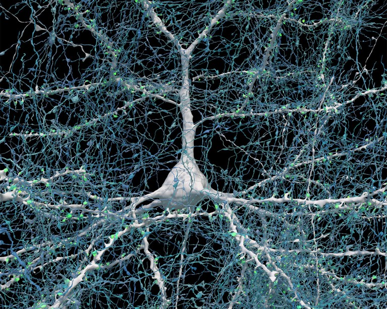
A single neuron (white) shown with 5,600 of the axons (blue) that connect to it. The synapses that make these connections are shown in green.Credit: Google Research & Lichtman Lab (Harvard University). Renderings by D. Berger (Harvard University)

When examining the model in detail, the researchers discovered unconventional neurons, including some that made up to 50 connections with each other. “In general, you would find a couple of connections at most between two neurons,” says Jain. Elsewhere, the model showed neurons with tendrils that formed knots around themselves. “Nobody had seen anything like this before,” Jain adds.

The team also found pairs of neurons that were near-perfect mirror images of each other. “We found two groups that would send their dendrites in two different directions, and sometimes there was a kind of mirror symmetry,” Jain says. It is unclear what role these features have in the brain.

Proofreaders needed

The map is so large that most of it has yet to be manually checked, and it could still contain errors created by the process of stitching so many images together. “Hundreds of cells have been ‘proofread’, but that’s obviously a few per cent of the 50,000 cells in there,” says Jain. He hopes that others will help to proofread parts of the map they are interested in. The team plans to produce similar maps of brain samples from other people — but a map of the entire brain is unlikely in the next few decades, he says.

“This paper is really the tour de force creation of a human cortex data set,” says Hongkui Zeng, director of the Allen Institute for Brain Science in Seattle. The vast amount of data that has been made freely accessible will “allow the community to look deeper into the micro-circuitry in the human cortex”, she adds.

Gaining a deeper understanding of how the cortex works could offer clues about how to treat some psychiatric and neurodegenerative diseases. “This map provides unprecedented details that can unveil new rules of neural connections and help to decipher the inner working of the human brain,” says Yongsoo Kim, a neuroscientist at Pennsylvania State University in Hershey.

[ad_2]

Source Article Link

Categories
Life Style

the dial in the brain that controls the immune system

[ad_1]

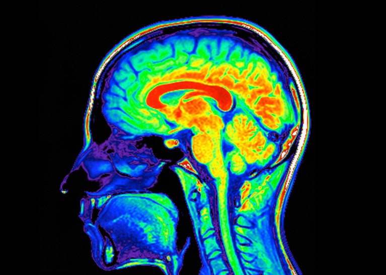
Coloured magnetic resonance imaging (MRI) scan of a sagittal section through a patient's head showing a healthy human brain and brain stem.

A population of neurons in the brain stem, the stalk-like structure that connects the bulk of the brain to the spinal cord, acts as the master dial for the immune system.Credit: Voisin/Phanie/Science Photo Library

Scientists have long known that the brain plays a part in the immune system — but how it does so has been a mystery. Now, scientists have identified cells in the brainstem that sense immune cues from the periphery of the body and act as master regulators of the body’s inflammatory response.

The results, published on 1 May in Nature1, suggest that the brain maintains a delicate balance between the molecular signals that promote inflammation and those that dampen it — a finding that could lead to treatments for autoimmune diseases and other conditions caused by an excessive immune response.

The discovery is akin to a black-swan event — unexpected but making perfect sense once revealed, says Ruslan Medzhitov, an immunologist at Yale University in New Haven, Connecticut. Scientists have known that the brainstem has many functions, such as controlling basic processes such as breathing. However, he adds, the study “shows that there is whole layer of biology that we haven’t even anticipated”.

The brain is watching

After sensing an intruder, the immune system unleashes a flood of immune cells and compounds that promote inflammation. This inflammatory response must be controlled with exquisite precision: if it’s too weak, the body is at greater risk of becoming infected; if it’s too strong, it can damage the body’s own tissues and organs.

Previous work has shown that the vagus nerve, a large network of nerve fibres that links the body with the brain, influences immune responses. However, the specific brain neurons that are activated by immune stimuli remained elusive, says Hao Jin, a neuroimmunologist at the US National Institute of Allergy and Infectious Diseases in Bethesda, Maryland, who led the work.

To investigate how the brain controls the body’s immune response, Jin and his colleagues monitored the activity of brain cells after injecting the abdomen of mice with bacterial compounds that trigger inflammation.

The researchers identified neurons in the brainstem that switched on in response to the immune triggers. Activating these neurons with a drug reduced the levels of inflammatory molecules in the mice’s blood. Silencing the neurons led to an uncontrolled immune response, with the number of inflammatory molecules increasing by 300% compared with the levels observed in mice with functional brainstem neurons. These nerve cells act as “a rheostat in the brain that ensures that an inflammatory response is maintained within the appropriate levels”, says study co-author Charles Zuker, a neuroscientist at Columbia University in New York City.

Further experiments revealed two discrete groups of neurons in the vagus nerve: one that responds to pro-inflammatory immune molecules and another that responds to anti-inflammatory molecules. These neurons relay their signals to the brain, allowing it to monitor the immune response as it unfolds. In mice with conditions characterized by an excessive immune response, artificially activating the vagal neurons that carry anti-inflammatory signals diminished inflammation.

Dampening autoimmune symptoms

Finding ways to control this newly discovered body–brain network would offer an approach to fixing broken immune responses in various conditions such as autoimmune diseases and even long COVID, a debilitating syndrome that can persist for years after a SARS-CoV-2 infection, Jin says.

There’s evidence that therapies targeting the vagus nerve can treat diseases such as multiple sclerosis and rheumatoid arthritis, suggesting that targeting the specific vagal neurons that carry immune signals might work in people, Zuker says. But, he cautions, “it’s a lot of work to go from here to there”.

Besides the neuronal network identified in the study, there might be other routes through which the body transmits immune signals to the brain, says Stephen Liberles, a neuroscientist at Harvard Medical School in Boston, Massachusetts. What’s more, the mechanisms by which the brain sends signals back to the immune system to regulate inflammation remain unclear. “We’re just scratching the surface,” he says. “We need to understand the rule book of how the brain and the immune system interact.”

[ad_2]

Source Article Link

Categories
Featured

‘Inspired by the human brain’: Intel debuts neuromorphic system that aims to mimic grey matter with a clear aim — making the machine exponentially faster and much more power efficient, just like us

[ad_1]

Neuromorphic computing is about mimicking the human brain’s structure to deliver more efficient data processing, including faster speeds and higher accuracy, and it’s a hot topic right now. A lot of universities and tech firms are working on it, including scientists at Intel who have built the world’s largest “brain-based” computing system for Sandia National Laboratories in New Mexico.

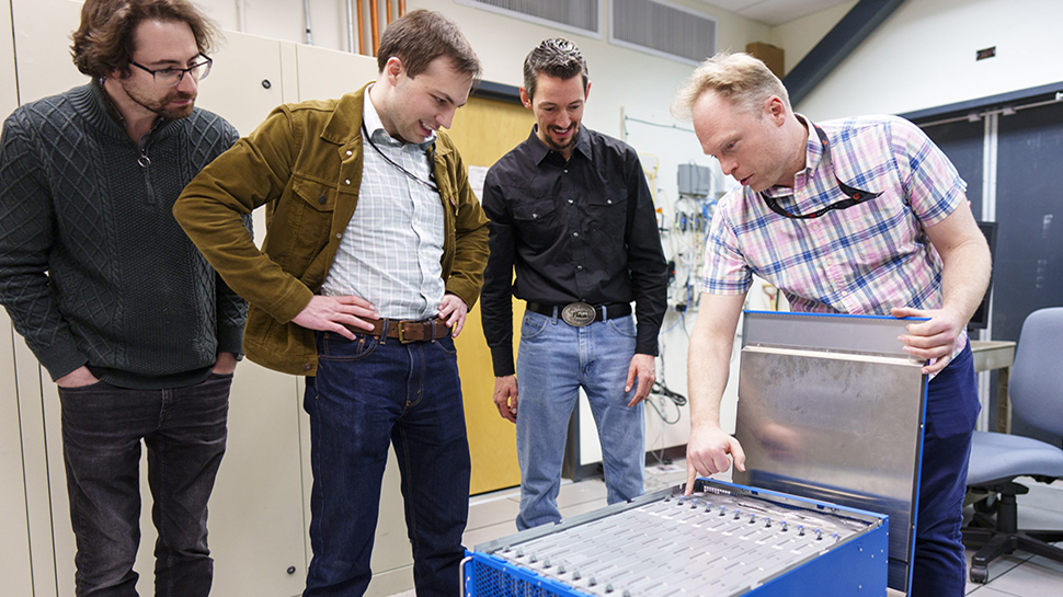
Intel’s creation, called Hala Point, is only the size of a microwave, but boasts 1.15 billion artificial neurons. That’s a massive step up from the 50 million neuron capacity of its predecessor, Pohoiki Springs, which debuted four years ago. There’s a theme with Intel’s naming in case you were wondering – they’re locations in Hawaii.

[ad_2]

Source Article Link

Categories
Life Style

Mini-colon and brain ‘organoids’ shed light on cancer and other diseases

[ad_1]

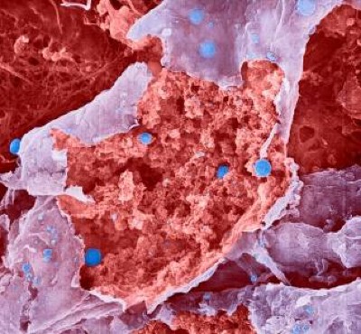
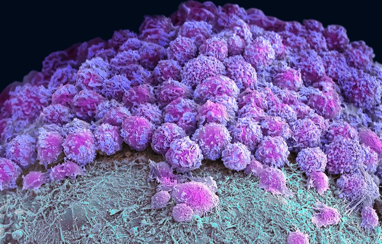
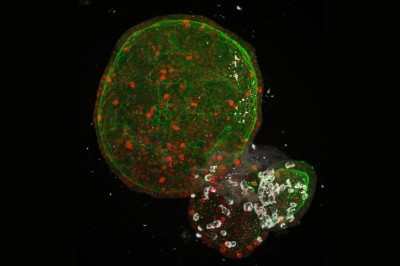
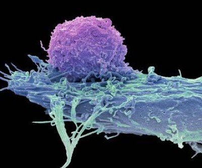
Coloured scanning electron micrograph (SEM) of a neural organoid.

Part of a brain organoid made using human stem cells (purple).Credit: Steve Gschmeissner/Science Photo Library

Researchers have hailed organoids — 3D clusters of cells that mimic aspects of human organs — as a potential way to test drugs and even eliminate some forms of animal experimentation. Now, in two studies published on 24 April in Nature1,2, biologists have developed gut and brain organoids that could improve understanding of colon cancer and help to develop treatments for a rare neurological disorder.

“In the last ten years, people spent a lot of time to develop and understand how to make organoids,” says Shuibing Chen, a chemical biologist at Weill Cornell Medical College in New York City. “But this is the time now to think more about how to use” the models.

Organoids — particularly those made from human stem cells — sometimes reveal things that animal models can’t, says Sergiu Pașca, a neuroscientist at Stanford University in California and a co-author of one of the studies1. Pașca’s group studies Timothy syndrome: a genetic disorder involving autism, neurological problems and heart conditions that affects only a few dozen people in the world. Timothy syndrome is caused by a single mutation in a gene called CACNA1C, which encodes a channel through which calcium ions enter cells including neurons.

Pașca says that there are no good animal models for Timothy syndrome because the underlying mutation doesn’t always cause the same symptoms in rodents. “It became very clear to us we’d need to find a way of testing in vivo,” he says.

Cultured cells

The researchers turned to brain organoids to recreate the disorder. They took stem cells from 3 people with the mutation that causes Timothy syndrome and cultured them for about 250 days, treating the cells with signalling molecules that encouraged them to turn into brain organoids containing every type of neuron found in the cerebral cortex, the outer layer of the brain. To create a more lifelike environment for the organoids, the team then injected these structures into the brains of rats, where the cells formed connections with the rodents’ own neurons. This made a system in which the researchers could test potential treatments for the disorder.

Human neurons have four different forms of this calcium channel, but only one of them is defective in Timothy syndrome. Getting rid of the mutated channel, the researchers suggest, would allow the other, healthy channels to take over.

To do this, they identified short pieces of nucleic acids called oligonucleotides that can stop cells producing the mutated form of the protein by interfering with genetic transcripts. Around two weeks after the researchers injected these oligonucleotides into the rats’ brains, most of the defective calcium channels in the organoids and the surrounding rat neurons had been replaced by other versions of the protein. The neurons in the organoids had also changed shape — from small, less complex forms similar to those in people with Timothy syndrome to larger, more complex shapes typical of healthy neurons. “To be honest, I didn’t think it would work so well,” Pașca says.

He adds that his group hope to test the therapy on people in clinical trials eventually, although they will first need to prove that the oligonucleotides are safe by testing them in non-human primates. The researchers think that the treatment would be effective for about three months, so people would need to receive frequent injections. The advantage, Pașca says, is that the biological effects of the treatment would be reversible and any side effects would be short-lived.

Miniature colon

In a separate paper2, bioengineer Matthias Lütolf at the Swiss Federal Institute of Technology in Lausanne and his colleagues used mouse stem cells to model tissue from the other end of the body: the gut tissue that makes up the colon and rectum. Organoids tend to grow in tight balls, so the researchers grew the cells on a scaffold to recreate the tube structures seen in real gut tissue.

To make a model of colon cancer, they engineered the cells to contain light-sensitive proteins attached to cancer-causing genes. This allowed them to use a blue laser to switch on the genes and trigger the growth of tumours at specific sites in the organoid, then watch how the tumours changed over the course of weeks.

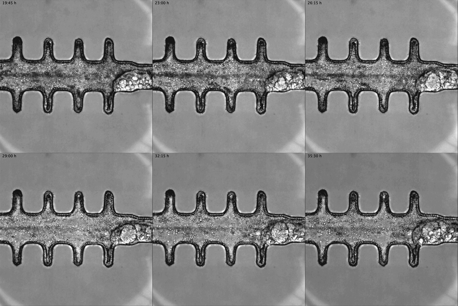
Animated sequence from a video showing mini-colon oncogenesis.

Researchers grew tumours inside a model of gut tissue created using mouse stem cells.Credit: Matthias P. Lutolf et al/Nature

When the researchers injected the cancerous cells into mice, the tumours looked similar to those seen in human colorectal cancer. The organoids accumulated fewer tumours when the researchers restricted calories in their medium, which also happens in people with colorectal cancer.

Lütolf was struck by the difference between the tumours triggered throughout the organoid. It would be difficult, he says, to use the organoids to screen a large number of new drugs, because of the differences and because they take a long time to produce. But he says organoids could be useful in investigating how drugs or immunotherapies kill tumours, and how factors such as a person’s environment and immune system affect the development and progression of colorectal cancer.

Lütolf and his group plan to manipulate the gut organoids to better reflect the human system, and hope that they could eventually replace animal models in some instances. Future organoid models, Lütolf says, could include bacteria that live in the gut or could be exposed to different levels of oxygen that mimic those available in various anatomical regions of the gut.

The two studies are “very well-designed”, Chen says. She adds that the organoid models seem particularly useful in showing the complexity of diseases such as Timothy syndrome and colorectal cancer, and how they progress over time. Now that researchers have found good ways to make organoids that model different organs, she says, the next steps will be learning how to scale up production for drug development, and making them more complex so that they reflect real human biology.

[ad_2]

Source Article Link

Categories
Life Style

Why queasiness kills hunger: brain circuit identified

[ad_1]

No one wants to eat when they have an upset stomach. To pinpoint exactly where in the brain this distaste for eating originates, scientists studied nauseated mice.

The work, published in Cell Reports on 27 March1, describes a previously uncharacterized cluster of brain cells that fire when a mouse is made to feel nauseous, but don’t fire when the mouse is simply full. This suggests that responses to satiety and nausea are governed by separate brain circuits.

“With artificial activation of this neuron, the mouse just doesn’t eat, even if it is super hungry,” says Wenyu Ding at the Max Planck Institute for Biological Intelligence in Martinsried, Germany, who led the study.

Ding and colleagues suspected that this group of neurons was involved in processing negative experiences, such as feeling queasy, so they injected the mice with a chemical that induces nausea and then scanned the animals’ brains. This confirmed that the neurons are active when mice feel nauseous.

Light bites

Using a light-based technique called optogenetics, the team artificially activated the neurons of mice that had been deprived of food in the hours before the experiment. When the neurons were ‘off’, the mice ate. When the researchers turned them on, the mice walked away mid-chow.

Researchers also blocked the activity of these neurons in nauseated mice that were hungry and found that the mice overcame their nausea to eat.

Understanding the brain circuits that control nausea is an important part of researching dysregulated eating, such as that seen in people with obesity and anorexia, says Haijiang Cai, a neuroscientist at the University of Arizona in Tucson.

A previous study2 described neurons near those characterized by the authors that also regulate eating, but that don’t differentiate between feelings of fullness and nausea. With their results, Ding and colleagues show that the two experiences are controlled by separate brain circuits.

“It’s going to be exciting in the future if we can target the neurocircuitry that controls satiation to suppress appetite, but not to cause nausea,” says Cai. For example, this information might assist in controlling the nausea caused by some appetite-suppressant drugs.

The same could be true in reverse, allowing someone to eat when they are nauseated. Nausea is a common side effect of many cancer treatments and makes it difficult for patients to stay properly nourished.

[ad_2]

Source Article Link

Categories
Life Style

COVID ‘brain fog’ linked to brain inflammation

[ad_1]

Hello Nature readers, would you like to get this Briefing in your inbox free every day? Sign up here.

Surgeons in protective clothing work on a patient.

Surgeons at Xijing Hospital in Xi’an, China, performed the first transplantation of a non-human liver into a human body.Credit: Xijing Hospital, Air Force Medical University in Xi’an China

Surgeons in China say they have transplanted a pig liver into a person’s body for the first time. With consent from the man’s family, the clinically dead patient received a liver from a pig that was genetically modified to prevent the recipient from rejecting the pig organ. The surgeons say the pig liver secreted more than 30 millilitres of bile every day, and the colour and texture of the liver remained normal after 10 days. In January, a US team conducted a similar experiment with a pig’s liver located outside a person’s body, and there have been further experiments with genetically modified pig kidneys and hearts.

Nature | 5 min read

Read more: Experts weigh in on the issues surrounding the xenotransplantation of pig organs in Nature Medicine (11 min read, from 2022)

Stellar detectives have identified seven stars that recently gobbled up a rocky planet. The planets seem to have been eaten during their stars’ relatively stable main-sequence period. If this is true, it means these systems have continued to be chaotic long after their formation, with planets disintegrating or falling into their star, says astronomer Johanna Teske. “It’s an inference at this point. We need to look at these systems in more detail.”

Nature | 3 min read

Reference: Nature paper

2,500

The number of researchers who have left Russia since it invaded Ukraine in February 2022, according to an estimate based on researchers’ ORCID identifiers. (Nature | 5 min read)

A slew of studies have identified how inflammation in the brains of people with COVID-19 might explain neurological symptoms such as loss of smell, headaches and memory problems. Growing evidence suggests that the immune response triggered by the virus leads indirectly to brain inflammation. One study found that people with long COVID and ‘brain fog’ had a leakier blood-brain barrier, which might let in molecules that cause inflammation.

Nature | 5 min read

Reference: Nature Neuroscience paper

Indian biotechnology company ImmunoACT is producing a much cheaper version of a cancer treatment known as chimeric antigen receptor (CAR) T-cell therapy. A single treatment of NexCAR19 costs between US$30,000 and $40,000 — a tenth of the price of comparable products now available. The safety profile also appears to be better than some US-approved CAR-T products. NexCAR19 is now being used to treat blood cancers in hospitals across India. “These are people for whom all other treatments have failed,” says immunologist Alka Dwivedi.

Nature | 6 min read

For 15 years, geoscientists have been involved in a complicated technical process to determine whether human impacts on Earth amount to a new geological epoch: the Anthropocene. This week, following a controversial vote and challenge, the final verdict has arrived from the International Union of Geological Sciences: we are not in a new epoch. The current lack of agreement on a start date should not detract from the Anthropocene as a concept, says a Nature editorial. The reality is that humans are leaving a discernible fingerprint on Earth systems.

Nature | 5 min read & Nature editorial | 5 min read

Features & opinion

AI image generators can amplify biased stereotypes in their output. There have been attempts to quash the problem by manual fine-tuning (which can have unintended consequences, for example generating diverse but historically inaccurate images) and by increasing the amount of training data. “People often claim that scale cancels out noise,” says cognitive scientist Abeba Birhane. “In fact, the good and the bad don’t balance out.” The most important step to understanding how these biases arise and how to avoid them is transparency, researchers say. “If a lot of the data sets are not open source, we don’t even know what problems exist,” says Birhane.

Nature | 12 min read

Invasive ant species such as the Argentine ant (Linepithema humile) have conquered the land so thoroughly that “it can seem as if the spread of global trade was an Argentine ant plot for world domination”, writes science journalist John Whitfield. He explores what makes these insects so successful, their effects on ecosystems and the temptation to compare their spread with humanity’s own power struggles. Ants “speak to life’s ability to escape our grasp, regardless of how we might try to order and exploit the world”, writes Whitfield. “There’s something hopeful about that, for the planet, if not for us.”

Aeon | 15 min read

Quote of the day

The psychoactive drug ketamine is increasingly being used to treat depression and other mood disorders, including by high-profile users such as entrepreneur Elon Musk or actor Matthew Perry. More than 40 clinical trials support its effectiveness in treating severe depression. But neuropsychopharmacologist David Nutt warns that those taking it need careful supervision. (Nature | 5 min read)

[ad_2]

Source Article Link

Categories
Computers

BMW’s Vision Neue Klasse X Has a Car-Wide Screen and a ‘Joy’ Brain

[ad_1]

“The ‘heart of joy’ effectively takes 30 years of experience and blends it into a single control unit,” BMW chief technical officer Frank Weber tells WIRED. “Everything that is driving-performance-related, chassis-control-related, powertrain-related—it’s all integrated into one control unit. If you love the idea of the ultimate driving machine, there are functions in there that are crazy. It’s the same for the infotainment system. To do it properly for your customers, you have to own the critical software stacks and the software development process.”

Crazy M Performance Promise

Weber insists that increasing the size of batteries is incompatible with BMW’s sustainability goals but promises authentic BMW vibes from its M-division high-performance derivatives. “Neue Klasse is ambitious and will do things far above what we have today,” he claims. “Future M cars will have close to a megawatt of power (1,340 bhp) with the ability to control each individual wheel.”

BMW's Vision Neue Klasse X driving on a desert road

Photograph: BMW

“Some people might miss the sound of a combustion engine but definitely not how the car behaves. It’s incredible. Everything required for M is baked into this new technology platform. As our engineers learned more about the capabilities of the system, so their confidence increased. It’s about how the car moves. And the control possibilities with electric cars means you can go crazy.”

Now back to the Vision X. If the iX and i7 are too much for you, then this new concept suggests a definite rebalancing of the aesthetic order. It’s a clean, modern looking car with a powerful but more nuanced sense of identity.

“We wanted to define the true-to-the-bone heritage of BMW,” head of i design Kai Langer tells me, “and the Vision X is our pure essence. Try to remove a line from this car and you just won’t be able to. The Vision X is clearly a BMW, even though it has completely different proportions. It’s uncomplicated, reduced, bold and alive.”

Getting 3D Grilled

BMW's Vision Neue Klasse X

Photograph: BMW

Interestingly, there are shades of the original 02 series, which debuted in 1966, as well as hints of the beloved ’70s 3.0 CSL, and even the early ’80s E30 3 series. The Vision X wears these influences lightly, but the fact that they’re there at all suggests that a rethink has occurred. The vertical double kidney grille will be reserved for BMW’s X SUV models henceforth, a subtle horizontal treatment being used on sedans and sports cars.

[ad_2]

Source Article Link

Categories
Life Style

CAR T cells can shrink deadly brain tumours — though for how long is unclear

[ad_1]

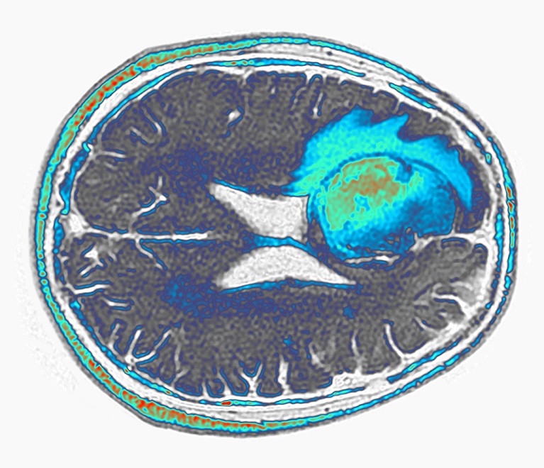
Coloured FLAIR (fluid-attenuated inversion recovery) magnetic resonance imaging scan (MRI) of an axia section through a human brain showing a glioblastoma affecting the frontal lobe.

A glioblastoma (green and blue, artificially coloured) grows in the frontal lobe of a person’s brain.Credit: Pr Michel Brauner, ISM/Science Photo Library

Two preliminary studies suggest that next-generation engineered immune cells show promise against one of the most feared forms of cancer.

A pair of papers published on 13 March, one in Nature Medicine1 and the other in New England Journal of Medicine2, describe the design and deployment of immune cells called chimeric antigen receptor T (CAR T) cells against glioblastoma, an aggressive and difficult-to-treat form of brain cancer. The average length of survival for people with this tumour is eight months.

Both teams found early hints of progress using CAR T cells that target two proteins made by glioblastoma cells, thereby marking those cells for destruction. CAR T cells are currently approved only for treating blood cancers such as leukaemia and are typically engineered to home in on only one target. But the new results add to mounting evidence that CAR T cells could be modified to treat a wider range of cancers.

“It lends credence to the potential power of CAR-T cells to make a difference in solid tumours, especially the brain,” says Bryan Choi, a neurosurgeon at Massachusetts General Hospital in Boston, and a lead author of the New England Journal of Medicine study. “It adds to the excitement that we might be able to move the needle.”

A highly lethal tumour

Glioblastomas offer a formidable challenge. Fast-growing glioblastomas can mix with healthy brain cells, forming diffuse tumours that are difficult to remove surgically. Surgery, chemotherapy and radiation therapy are typically the only treatment options and tend to produce short-lived, partial responses.

In CAR-T therapy, a person’s own T cells are removed from the body and kitted out with proteins that help the cells home in on tumours. The souped-up cells are then reinfused into the body.

In the past few years, researchers have been developing CAR T cells that target specific molecules made by some glioblastomas. The two new papers take this a step farther by designing CAR T cells that target not one type of molecule but two.

In one approach, Choi and his colleagues designed CAR T cells to latch onto a mutated form of a protein called EGFR that is produced by some glioblastomas. The CAR T cells also secreted antibodies that bind to both T cells and the unmutated form of EGFR, which is not typically made by brain cells but is often made by glioblastoma cells. The result is a CAR-T therapy that unleashes the immune system against cells that express either the mutated or the unmutated form of EGFR.

Choi and his team administered these cells to three adults with glioblastoma. Tumours appeared to shrink in all three, but later recurred. One man who received the treatment, however, had a response that lasted for more than six months.

Seven months and counting

The other team, led by Stephen Bagley, a neurooncologist at the University of Pennsylvania Perelman School of Medicine in Philadelphia, used CAR T cells that target both EGFR and another protein found in glioblastomas called interleukin-13 receptor alpha 2. Tumours appeared to shrink in all six of the people they treated. One participant’s glioblastoma began to grow again within a month, but one participant has not shown signs of tumour progression for seven months so far, says Bagley. Of the remaining four participants, one left the trial, and tumours have not rebounded in the remaining three, but they are within six months of treatment.

The results are promising, but the goal is to generate longer-lasting responses, says Bagley. It was exciting, he says, to watch tumours shrink in the first day after CAR-T therapy. “We hadn’t seen that before,” he says. “We were thrilled.”

But the excitement faded as participants relapsed after treatment: “It’s very humbling to go on that roller coaster ride,” he says. “One week you feel like you’ve made a real difference in their lives, and the next week the tumour is back again.”

Versatile T cells

The field will eagerly await additional results, says Sneha Ramakrishna, a paediatric oncologist at Stanford Medicine in California. The size of glioblastomas is notoriously difficult to measure because of their diffuse shape, and apparent changes in tumour size could be affected by inflammation following surgery to administer the CAR T cells directly into the brain.

But the images are impressive, and measures of tumour RNA in Choi’s study suggest that the tumours might have indeed shrunk, says Ramakrishna. And constructing CAR T cells with multiple targets could ultimately yield long lasting therapies, she says, by making it more difficult for cancer cells to develop ways to resist the therapy.

“I’m looking forward to seeing what they do over time,” she says. “I hope that as we get more experience, we can learn how to make the right CAR for our patients.”

[ad_2]

Source Article Link

Categories
Politics

How the brain coordinates speaking and breathing

[ad_1]

MIT researchers have discovered a brain circuit that drives vocalization and ensures that you talk only when you breathe out, and stop talking when you breathe in.

The newly discovered circuit controls two actions that are required for vocalization: narrowing of the larynx and exhaling air from the lungs. The researchers also found that this vocalization circuit is under the command of a brainstem region that regulates the breathing rhythm, which ensures that breathing remains dominant over speech.

“When you need to breathe in, you have to stop vocalization. We found that the neurons that control vocalization receive direct inhibitory input from the breathing rhythm generator,” says Fan Wang, an MIT professor of brain and cognitive sciences, a member of MIT’s McGovern Institute for Brain Research, and the senior author of the study.

Jaehong Park, a Duke University graduate student who is currently a visiting student at MIT, is the lead author of the study, which appears today in Science. Other authors of the paper include MIT technical associates Seonmi Choi and Andrew Harrahill, former MIT research scientist Jun Takatoh, and Duke University researchers Shengli Zhao and Bao-Xia Han.

Vocalization control

Located in the larynx, the vocal cords are two muscular bands that can open and close. When they are mostly closed, or adducted, air exhaled from the lungs generates sound as it passes through the cords.

The MIT team set out to study how the brain controls this vocalization process, using a mouse model. Mice communicate with each other using sounds known as ultrasonic vocalizations (USVs), which they produce using the unique whistling mechanism of exhaling air through a small hole between nearly closed vocal cords.

“We wanted to understand what are the neurons that control the vocal cord adduction, and then how do those neurons interact with the breathing circuit?” Wang says.

To figure that out, the researchers used a technique that allows them to map the synaptic connections between neurons. They knew that vocal cord adduction is controlled by laryngeal motor neurons, so they began by tracing backward to find the neurons that innervate those motor neurons.

This revealed that one major source of input is a group of premotor neurons found in the hindbrain region called the retroambiguus nucleus (RAm). Previous studies have shown that this area is involved in vocalization, but it wasn’t known exactly which part of the RAm was required or how it enabled sound production.

The researchers found that these synaptic tracing-labeled RAm neurons were strongly activated during USVs. This observation prompted the team to use an activity-dependent method to target these vocalization-specific RAm neurons, termed as RAmVOC. They used chemogenetics and optogenetics to explore what would happen if they silenced or stimulated their activity. When the researchers blocked the RAmVOC neurons, the mice were no longer able to produce USVs or any other kind of vocalization. Their vocal cords did not close, and their abdominal muscles did not contract, as they normally do during exhalation for vocalization.

Conversely, when the RAmVOC neurons were activated, the vocal cords closed, the mice exhaled, and USVs were produced. However, if the stimulation lasted two seconds or longer, these USVs would be interrupted by inhalations, suggesting that the process is under control of the same part of the brain that regulates breathing.

“Breathing is a survival need,” Wang says. “Even though these neurons are sufficient to elicit vocalization, they are under the control of breathing, which can override our optogenetic stimulation.”

Rhythm generation

Additional synaptic mapping revealed that neurons in a part of the brainstem called the pre-Bötzinger complex, which acts as a rhythm generator for inhalation, provide direct inhibitory input to the RAmVOC neurons.

“The pre-Bötzinger complex generates inhalation rhythms automatically and continuously, and the inhibitory neurons in that region project to these vocalization premotor neurons and essentially can shut them down,” Wang says.

This ensures that breathing remains dominant over speech production, and that we have to pause to breathe while speaking.

The researchers believe that although human speech production is more complex than mouse vocalization, the circuit they identified in mice plays the conserved role in speech production and breathing in humans.

“Even though the exact mechanism and complexity of vocalization in mice and humans is really different, the fundamental vocalization process, called phonation, which requires vocal cord closure and the exhalation of air, is shared in both the human and the mouse,” Park says.

The researchers now hope to study how other functions such as coughing and swallowing food may be affected by the brain circuits that control breathing and vocalization.

The research was funded by the National Institutes of Health.

[ad_2]

Source Article Link