US President Joe Biden signed legislation last month that could trigger a nationwide ban of the popular social-media app TikTok within a year. Researchers who use the app to communicate science to curious followers, study social trends and earn money to support themselves are dismayed and frustrated.
Creating YouTube and TikTok videos is improving my lab leadership
TikTok is owned by ByteDance, a firm based in Beijing, China — which, amid growing US–China tensions, has raised national-security concerns among US officials, related to the Chinese government’s access to user data. On 7 May, TikTok filed a lawsuit calling the legislation, which gives ByteDance nine months to a year to find a US-based buyer for the app, an “extraordinary intrusion on free speech rights”. If the ban goes into effect, users in the United States will no longer be able to add the app to their devices or install updated versions.
Morgan Johnston, a neuroscientist at the University of Texas at San Antonio, worries that young people who use TikTok will lose an outlet to learn about science and find community. Sixty-three per cent of US teenagers aged 13 to 17 and 33% of US adults use the app, according to several surveys conducted last year by the Pew Research Center in Washington DC and market-research firm Ipsos in New York City. Although dance routines and pop-culture discussions are often trending on the app, many people use it to learn about science, says Johnston, who runs the account @askaneuroscientist. She posts videos about her research on the impact of stress on learning and answers questions from her 37,600 followers. “I love the interaction part of it,” she says.
Nature spoke to five scientists and communicators in the United States about what they will do if the ban goes into effect. Most of them acknowledge the data-security concerns, but say that the legislation would cut off a thriving platform for science education and outreach, especially among young people who are seeking information from trusted sources.
“We need stricter laws on what data can be collected and sold, but this legislation doesn’t do that,” says Johnston, who uses her channel to talk about her mental-health journey as she navigates graduate school as a first-generation PhD student from a rural area. “My following is young adults who are in the process of making their career decisions — and they’re really curious.”
Engaged audiences
The seismic growth of the app during lockdowns initiated because of the COVID-19 pandemic helped to make TikTok a “massive platform for outreach”, says Jamie Zaccaria, a media and outreach specialist at the Ocean Exploration Trust, a research-focused non-profit organization in New London, Connecticut. In 2022, the trust launched its TikTok account, @nautiluslive, which streams footage of deep-sea expeditions narrated in real time by excited researchers discovering striking marine creatures. The account has more than half a million followers, and some ask for educational resources or advice on how to pursue a career in ocean science.
Researchers with the Ocean Exploration Trust marvel at a mysterious deep-sea jellyfish in this video (shown here on YouTube, but also available on TikTok).
The Ocean Exploration Trust declined to comment on the legislation, but it has several successful social-media accounts, including on YouTube, Facebook and Instagram, to which it could shift its focus if the ban goes into effect.
Some content creators with smaller online followings might not have that option. Michael Rhodes, a neuroscientist at Saint Vincent College in Latrobe, Pennsylvania, started posting on his TikTok account, @rhodeslovesneuroscience, in 2020 to boost morale among undergraduate students in his classes. With their help and with help from researchers in his lab, Rhodes posts educational videos about anatomy, physiology and pharmacology, including dance routines that demonstrate the actions performed by specific muscle groups and skits that explain how drugs work. “I’ve become a better professor because it makes me take a step back and look at information differently,” he says.
Rhodes says that most of his roughly 189,000 followers are students or early-career health-care professionals. But with all his academic responsibilities, he isn’t sure that he will be able to pivot to a new platform if TikTok disappears in the United States. “If a third of the country is using TikTok, that should tell you something about its popularity,” he adds.
Secret sauce
One thing that makes TikTok different from other social-networking apps is how its algorithm curates content from across the platform — not just the accounts a user follows — to appeal to the specific interests of each user, says Matt Motta, a health-communications researcher at Boston University’s School of Public Health. “That becomes a way for scientists to have their messages transmitted to audiences that may not self-select into them,” he says.
TikTok’s critics say that this proprietary algorithm imbues the app with addictive properties that can drive the spread of misinformation and contribute to the US mental-health crisis. But, recognizing its reach, Motta and his colleagues are studying how TikTok could be harnessed for good by training mental-health content creators on the app to disseminate evidence-based information among their followers1.
“It’s important to remember that some scientists are working with TikTok to study social phenomena. And if TikTok were to go away, our ability to do that would be significantly hindered,” Motta says. At the same time, Motta and others acknowledge concerns about data security related to the use of social-media apps such as TikTok.
The TikTok creator and organic chemist known as Chem Thug explains why batteries bounce when they run out of juice (shown here on YouTube, also available on TikTok).
Digital privacy is part of the reason that the organic-chemistry PhD student behind the viral @chem.thug TikTok account does not share his real identity or the university that he attends. His conversational explainer videos put scientific concepts in a real-world context for more than 284,300 followers on TikTok and around 10,000 on YouTube. For example, in one of his popular clips about household chemicals, Chem Thug explains why zinc-based batteries become “bouncy” as they lose charge. “I think everybody’s life is enriched by a better, deeper understanding of chemistry,” he says. Like Johnston, Chem Thug has monetized his account as a supplemental line of income to support himself during graduate school.
Chem Thug is cautious about putting his personal information on the Internet, but says that he doesn’t “see Bytedance as being any more nefarious than any other large corporation with interest in making as much money as possible”.
Few sources who spoke to Nature anticipate that a ban will go into effect on the proposed timeline, especially considering that the lawsuit filed by TikTok will undoubtedly tie up the legislation in courts. But the spectre of the ban is sparking serious conversations among TikTok scientists, especially those who sought refuge on the app after billionaire Elon Musk bought the social-media platform Twitter (now X) and made many unpopular changes. “Where are we going to recreate this community?” Johnston asks. “There’s not really a consensus.”
Hello Nature readers, would you like to get this Briefing in your inbox free every day? Sign up here.
Zhang Yongzhen, the first person to publicly release the genome sequence of the virus that causes COVID-19, was camping outside his laboratory.Credit: Dake Kang/AP via Alamy
Noted Chinese virologist Zhang Yongzhen appears to be back at work following a dispute that saw him sleeping in the street outside his own lab. According to social media posts on Zhang’s Weibo account, his group was given two days to relocate to a new lab that lacked sufficient biosafety controls. In 2020, Zhang and long-time collaborator Edward Holmes, a virologist in Australia, were first to publicly release the genome of SARS-CoV-2 — a choice credited as key to the swift development of COVID-19 vaccines. But Zhang’s research output has since dwindled, which Holmes blames on an effort to sideline Zhang for unauthorized sharing of data. “It is heartbreaking to watch,” he says. “It is unfathomable to me to have a scientist of that calibre sleeping outside his lab.”
A ‘challenge trial’ early in the COVID-19 pandemic that aimed to infect 35 volunteers on purpose to study treatments ended after none of them got sick, a paper detailing the results has revealed. Fourteen of the participants then caught the Omicron strain after being released from quarantine. The strains used in challenge trials are produced under stringent conditions — a process that can take months. This can put them well out-of-date compared to emerging variants that can overcome widespread immunity. “We need a challenge strain that’s more representative of what’s circulating in the community,” says vaccine scientist Anna Durbin.
India’s leading social-science research institute, the Centre for Policy Research (CPR), is reeling after a January decision by the government — currently being challenged in court — banned it from taking money from international funders, including the Bill & Melinda Gates Foundation and the UK Foreign, Commonwealth & Development Office. The tax authorities also levied the institution with a 10 crore rupees (US$1.2 million) bill.
In an opinion article, former CPR chief executive Yamini Aiyar, who stepped down in March, says this is part of a pattern of attacks on institutions conducting independent research. “The documented drop in academic freedom is part of a broader decline in India’s vibrant culture of public debate,” she writes. “At a juncture when critical feedback and effective consultation are required to secure the country’s long-term growth and prosperity… it has now become increasingly common for technocrats in government to seek to discredit researchers and suppress research.”
Scientists are racing to find out whether chimeric antigen receptor (CAR) T-cell therapies, one of the most celebrated new cancer treatments in decades, could be causing new malignancies. The US Food and Drug Administration received 33 reports of lymphomas among some 30,000 people who had been treated. It remains unclear how many, if any, of the new cancers came from the CAR T cells or from other therapies the patients had received. “Most cancer therapies can cause cancer. This is one of the paradoxes of our business,” says paediatric oncologist Crystal Mackall.
The Netflix series 3 Body Problem is a hit — but is the mind-bending tale of a group of alien-battling Oxford physicists good science? Nature asked Xavier Dumusque, a planetary scientist who has studied the three-star system Alpha Centauri, Younan Xia, a materials scientist who has worked with cutting-edge nanotechnologies and Matt Kenzie, a particle physicist and the scientific adviser for the show.
Like many regions, Africa faces challenges to infrastructure, institutions and ecosystems. “But the current circumstances also offer an opportunity for African nations,” argues agricultural economist Alfred Bizoza. “Despite — or perhaps because of — its challenges, Africa is already a hub for sustainable innovation.” He calls for ‘supported independence’ of science and innovation in the continent, with the aim of designing innovations that work for African researchers and African people.
The mystery of consciousness and the quirks of quantum physics are signs of a blind spot at the heart of science, write astronomer Adam Frank, theoretical physicist Marcelo Gleiser and philosopher Evan Thompson in their new book of the same name. (Big Think | 6 min read — or read a review of the book in Science, 5 min read)
A couple of weeks ago, we told you (in a story about left- and right-handed molecules) that the drug thalidomide showed to tragic effect why it’s important to separate molecules’ mirror-image forms: one version is a sedative, the other causes congenital disabilities when taken during pregnancy.
While technically true — the different versions do have different effects — Editorial Director of the Physics & Chemistry Nature journals (and former editor of Nature Chemistry) Stuart Cantrill alerted us that the real-world implications have become something of a chemistry urban myth. “In the body, the two forms will interconvert,” notes Cantrill. “So even if you give the ‘safe’ mirror image form it will convert into the version that is not safe (well, you’ll get a 50:50 mixture) and biological studies confirm that it leads to embryonic defects just as if you gave the mixed versions in the first place.” The persistence of the tale was explored in detail in Nature Chemistry in 2010 by chemist Michelle Francl (who blew our minds in January with her revelation that the secret to a great cup of tea is a pinch of salt).
Want more? Sign up to our other free Nature Briefing newsletters:
• Nature Briefing: Microbiology — the most abundant living entities on our planet — microorganisms — and the role they play in health, the environment and food systems.
When Bente Klarlund Pedersen wakes up in the morning, the first thing she does is pull on her trainers and go for a 5-kilometre run — and it’s not just about staying fit. “It’s when I think and solve problems without knowing it,” says Klarlund Pedersen, who specializes in internal medicine and infectious diseases at the University of Copenhagen. “It’s very important for my well-being.”
Whether it’s running or lifting weights, it’s no secret that exercise is good for your health. Research has found that briskly walking for 450 minutes each week is associated with living around 4.5 years longer than doing no leisure-time exercise1, and that engaging in regular physical activity can fortify the immune system and stave off chronic diseases, such as cancer, cardiovascular disease and type 2 diabetes. But, says Dafna Bar-Sagi, a cell biologist at New York University, the burning question is how does exercise deliver its health-boosting effects?
“We know that it is good, but there is still a huge gap in understanding what it is doing to cells,” says Bar-Sagi, who walks on a treadmill for 30 minutes, five days a week.
In the past decade, researchers have started to build a picture of the vast maze of cellular and molecular processes that are triggered throughout the body during — and even after — a workout. Some of these processes dial down inflammation, whereas others ramp up cellular repair and maintenance. Exercise also prompts cells to release signalling molecules that carry a frenzy of messages between organs and tissues: from muscle cells to the immune and cardiovascular systems, or from the liver to the brain.
But researchers are just beginning to work out the meaning of this cacophony of crosstalk, says Atul Shahaji Deshmukh, a molecular biologist at the University of Copenhagen. “Any single molecule doesn’t work alone in the system,” says Deshmukh, who enjoys mountain biking during the summer. “It’s an entire network that functions together.”
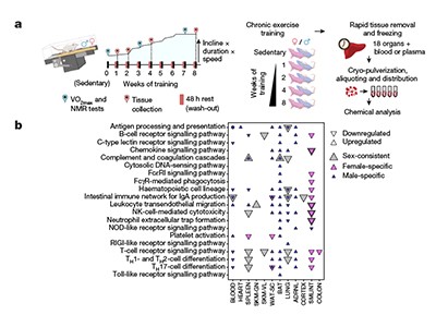
Endurance exercise causes a multi-organ full-body molecular reaction
Exercise is also attracting attention from funders. The US National Institutes of Health (NIH), for instance, has invested US$170 million into a six-year study of people and rats that aims to create a comprehensive map of the molecules behind the effects of exercise, and how they change during and after a workout. The consortium behind the study has already published its first tranche of data from studies in rats, which explores how exercise induces changes across organs, tissues and gene expression, and how those changes differ between sexes2–4.
Building a sharper view of the molecular world of exercise could reveal therapeutic targets for drugs that mimic its effects — potentially offering the benefits of exercise in a pill. However, whether such drugs can simulate all the advantages of the real thing is controversial.
The work could also offer clues about which types of physical activity can benefit people with chronic illnesses, says Klarlund Pedersen. “We think you can prescribe exercise as you can prescribe a medicine,” she says.
Hard-wired for exercise
Exercise is a fundamental thread in the human evolutionary story. Although other primates evolved as fairly sedentary species, humans switched to a hunter-gatherer lifestyle that demanded walking long distances, carrying heavy loads of food and occasionally running from threats.
Those with better athletic prowess were better equipped to live longer lives, which made exercise a core part of human physiology, says Daniel Lieberman, a palaeoanthropologist at Harvard University in Cambridge, Massachusetts. The switch to a more active lifestyle led to changes in the human body: exercise burns up energy that would otherwise be stored as fat, which, in excess amounts, increases the risk of cardiovascular disease, type 2 diabetes and some cancers. The stress induced by running or pumping iron has the potential to damage cells, but it also kick-starts a cascade of cellular processes that work to reverse those effects. This can leave the body in better shape than it would be without exercise, says Lieberman.
Researchers have been exploring some of the biological changes that occur during exercise for more than a century. In 1910, pharmacologist Fred Ransom at the University of Cambridge, UK, discovered that skeletal muscle cells secrete lactic acid, which is created when the body breaks down glucose and turns it into fuel5. And in 1961, researchers speculated that skeletal muscle releases a substance that helps to regulate glucose during exercise6.
More clues were in store. In 1999, Klarlund Pedersen and her colleagues collected blood samples from runners before and after they took part in a marathon and found that several cytokines — a type of immune molecule — spiked immediately after exercise and that many remained elevated for up to 4 hours afterwards7. Among these cytokines were interleukin-6 (IL-6), a multifaceted protein that is a key player in the body’s defence response. The following year, Klarlund Pedersen and her colleagues discovered8 that IL-6 is secreted by contracting muscles during exercise, making it an ‘exerkine’ — the umbrella term for compounds produced in response to exercise.
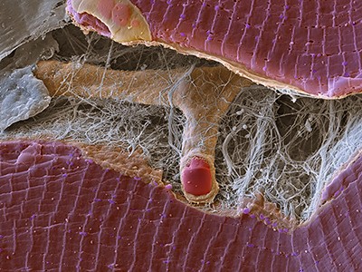
Exercising regularly can strengthen the immune system and stave off disease.Credit: Mike Kemp/Getty
High levels of IL-6 can be beneficial or harmful, depending on how it is provoked. At rest, too much IL-6 has an inflammatory effect and is linked to obesity and insulin resistance, a hallmark of type 2 diabetes, says Klarlund Pedersen. But when exercising, the molecule activates its more calming family members, such as IL-10 and IL-1ra, which tone down inflammation and its harmful effects. “With each bout of exercise, you provoke an anti-inflammatory response,” says Klarlund Pedersen. Although some physical activity is better than none, high-intensity, long-duration exercise that engages large muscles — such as running or cycling — will crank up IL-6 production, adds Klarlund Pedersen.
Exercise is a balancing act in other ways, too. Physical activity produces cellular stress, and certain molecules counterbalance this damaging effect. When mitochondria — the powerhouses that supply energy in cells — ramp up production during exercise, they also produce more by-products called reactive oxygen species (ROS), which, in excessive amounts, can damage proteins, lipids and DNA. But these ROS also kick-start a horde of protective processes during exercise, offsetting their more toxic effects and fortifying cellular defences.
Among the molecular stars in this maintenance and repair arsenal are the proteins PGC-1α, which regulates important skeletal muscle genes, and NRF2, which activates genes that encode protective antioxidant enzymes. During exercise, the body has learnt to benefit from a fundamentally stressful process. “If stress doesn’t kill you it makes you stronger,” says Ye Tian, a geneticist at the Institute of Genetics and Developmental Biology, Chinese Academy of Sciences, Beijing.
Exerkines everywhere
Since IL-6 ushered in the exerkine era, the explosion of multiomics — an approach that combines various biological data sets, such as the proteome and metabolome — has allowed researchers to go beyond chasing single molecules. They can now begin untangling the convoluted molecular web that lies behind exercise, and how it interacts with different systems across the body, says Michael Snyder, a geneticist at Stanford University in California, who recently switched from running to weightlifting. “We need to understand how these all work together, because [humans] are a homeostatic machine that needs to be properly tuned,” he says.
In 2020, Snyder and his colleagues took blood samples from 36 people aged between 40 and 75 years old before, during and at various time intervals after the volunteers ran on a treadmill. The team used multiomic profiling to measure more than 17,000 molecules, more than half of which showed significant changes after exercise9. They also found that exercise triggered an elaborate ‘choreography’ of biological processes such as energy metabolism, oxidative stress and inflammation. Creating a catalogue of exercise molecules is an important first step in understanding their effects on the body, says Snyder.
How an exercise habit paves the way for injured muscles to heal
Other studies have probed how exercise affects cell types. A 2022 study in mice led by Jonathan Long, a pathologist at Stanford University, identified more than 200 types of protein that were expressed differently by 21 cell types in response to exercise10. The researchers were expecting to find that cells in the liver, muscle and bone would be most sensitive to exercise, but to their surprise, they found that a much more widespread type of cell, one that appears in many tissues and organs, showed the biggest changes in the proteins that it cranked out or turned down. The findings suggest that more cell types shift gears during a workout than was previously thought, although what these changes mean for the body is still an open question, says Long.
The findings also showed that after exercise, the mice’s liver cells squeezed out several types of carboxylesterase enzyme, which are known to ramp up metabolism. When Long and his colleagues genetically tweaked mice so that their livers expressed elevated levels of these metabolism-enhancing enzymes, and then fed them a diet of fatty foods, the mice didn’t gain weight. They also had increased endurance when they ran on a treadmill. “The improvement in exercise performance by these secreted carboxylesterases was not known before,” says Long, whose weekly exercise regime involves swimming and lifting weights. He adds that if the enzymes could be produced in the right quantities and purity, they could possibly be used as exercise-mimicking compounds.
During a workout, distant organs and tissues communicate with each other through molecular signals. Along with exerkines, extracellular vesicles (EVs) — nanosized, bubble-shaped structures that carry biological material — could be one of the mechanisms behind organ and tissue crosstalk, says Mark Febbraio, a former triathlete who is now an exercise physiologist at Monash University in Melbourne, Australia. In 2018, Febbraio and his team inserted tubes into the femoral arteries of 11 healthy men and drew blood before and after they rode an exercise bike at an increasing pace for an hour. During and after exercise, but not at rest, they found a spike in the levels of more than 300 types of protein that compose or are carried by EVs11.
When the team then collected EVs from mice that had run on a treadmill and injected them into another group of healthy mice, most of the EVs ended up in liver cells. In a separate mouse study that is yet to be published, Febbraio and his colleagues found hints that the contents of these liver-bound EVs can arrest a type of liver disease. A big question is whether EVs also deposit genetic material into different cells, and if so, what that means for the body. “We still don’t know a great deal,” he says.
Exercise as medicine
Larger efforts are under way to build a detailed molecular snapshot of how exercise exerts its health-boosting effects across tissues and organs. In 2016, the NIH established the Molecular Transducers of Physical Activity Consortium (MoTrPAC), a six-year study on around 2,600 people and more than 800 rats that aims to generate a molecular map of exercise. The effort — one of the largest studies on physical activity — is teasing apart the effects of aerobic and endurance exercise on multiple tissue types across different ages and fitness levels.
The first data set is from rats that completed one to eight weeks of treadmill training, and had blood and tissue samples collected at the end. The researchers pinpointed thousands of molecular changes throughout the rats’ bodies, many of which could have a protective effect on health, such as dialling down inflammatory bowel disease and tissue injury2. A separate study3 found that the effects of endurance training differed across sexes: markers associated with the breakdown of fat increased in male fat tissue, driving fat loss, whereas female fat tissue showed an increase in markers related to fat-cell maintenance and insulin signalling, which might protect against cardiometabolic diseases. A third study4 found that exercise alters the expression of genes linked to diseases such as asthma, and could help to trigger similar adaptive responses.
Focus on exercise metabolism and health
A big goal is to uncover why exercise has such varied effects on people of different sexes, ages and ethnic backgrounds, says Snyder, who is a member of the MoTrPAC team. “It’s very obvious that some people benefit better than others,” he says.
Researchers hope that the reams of molecular data will eventually help clinicians to develop tailored exercise prescriptions for people with chronic diseases, says MoTrPAC team member Bret Goodpaster, an exercise physiologist at the University of Pittsburgh in Pennsylvania. Farther down the track, such insights could be used to develop therapeutics that mimic some of the beneficial effects of exercise in people who are too ill to work out, he says. “That’s not to say that we will have exercise in a pill, but there are certain aspects of exercise that could be druggable,” says Goodpaster, who has taken part in triathlons, marathons and cycling races.
Several teams are already in the early stages of developing exercise-mimicking therapeutics. In March 2023, a team led by Thomas Burris, a pharmacologist at the University of Florida in Gainesville, identified a compound that targets proteins called oestrogen-related receptors, which are known to trigger key metabolic pathways in energy-intensive tissues, such as heart and skeletal muscle, particularly during exercise12. When the researchers administered the compound — called SLU-PP-332 — to mice, they found that the treated rodents were able to run 70% longer and 45% farther than untreated mice. Six months later, a separate study, also led by Burris, found that obese mice treated with the drug lost weight and gained less fat than those that didn’t receive the treatment — even though their diet was the same and they didn’t exercise any more than usual13.
There is already evidence that exercise itself acts like medicine. In 2022, Bar-Sagi and her colleagues found that mice with pancreatic cancer had elevated levels of CD8 T cells — which destroy cancerous and virus-infected cells — when they did 30 minutes of aerobic exercise for 5 days a week14. These killer cells express a receptor for IL-15, another exerkine released by muscles during exercise. The researchers found that when CD8 T cells bind to IL-15, they unleash a more powerful immune response on tumours in the pancreas. This effect prolonged survival of mice with tumours by around 40%, compared with that of control mice. The findings held up when Bar-Sagi and her team analysed tumour tissue taken from people with pancreatic cancer. Those who did 60 minutes of aerobic and strength training each week had more CD8 T cells, and were twice as likely to survive for up to 5 years, than were people in the control group.
Although exercising more is a no-brainer for improving health, around 25% of adults globally do not meet the World Health Organization’s recommended levels of exercise each week: 150–300 minutes or more of moderate-intensity exercise, such as a brisk walk; or 75–150 minutes of vigorous-intensity exercise, such as running. David James, an exercise physiologist at the University of Sydney in Australia, who rides his bike to work each day, says that understanding the inner workings of exercise could help to develop clearer public-health messages about why physical activity is important and how it can offset the risk of getting chronic diseases. “That’s a powerful message,” says James.
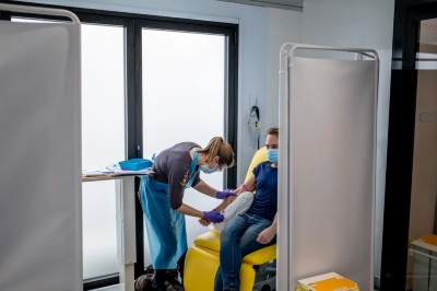
When Paul Zimmer-Harwood volunteered to be intentionally infected with SARS-CoV-2, he wasn’t sure what to expect. He was ready for a repeat of his first brush with COVID-19, through a naturally acquired infection that gave him influenza-like symptoms. But he hoped his immunity would help him feel well enough to use the indoor bicycle trainer that he had brought into quarantine.
It turned out that Zimmer-Harwood, a PhD student at University of Oxford, UK, had nothing to worry about. Neither he nor any of the 35 other people who participated in the ‘challenge’ trial actually got COVID-19.
Scientists deliberately gave people COVID — here’s what they learnt
The study’s results, published on 1 May in Lancet Microbe1, raise questions about the usefulness of COVID-19 challenge trials for testing vaccines, drugs and other therapeutics. “If you can’t get people infected, then you can’t test those things,” says Tom Peacock, a virologist at Imperial College London. Viral strains used in challenge trials take many months to produce, making it impossible to match emerging circulating variants that can overcome high levels of existing immunity in populations.
Researchers use challenge trials to understand infections and quickly test vaccines and therapies. In March 2021, after months of ethical debate, UK researchers launched the world’s first COVID-19 challenge trial. The study2 identified a minuscule dose of the SARS-CoV-2 strain that circulated in the early days of the pandemic that could infect about half of the participants, who had not previously been infected with the virus (at that time, vaccines weren’t yet widely available).
In parallel, a team led by Helen McShane, an infectious-disease researcher at Oxford, launched a second SARS-CoV-2 challenge study in people — including Zimmer-Harwood — who had recovered from naturally caught SARS-CoV-2 infections, caused by a range of variants. The trial later enrolled participants who had also been vaccinated.
Evolving strains
The first participants got the same tiny dose of the ‘ancestral’ SARS-CoV-2 strain as did those in the first trial. When nobody developed a sustained infection, the researchers increased the dose by more and more in subsequent groups of participants, until they reached a level 10,000 times the initial dose. A few volunteers developed short-lived infections, but these quickly vanished.
These volunteers want to be infected with disease to aid research — will their altruism help?
“We were quite surprised,” says Susan Jackson, a study clinician at Oxford and co-author of the latest study. “Moving forward, if you want a COVID challenge study, you’re going to have to find a dose that infects people.”
An ongoing COVID-19 challenge trial at Imperial College London, in which participants have been exposed to the Delta SARS-CoV-2 variant, has also encountered problems with infecting participants reliably, says Christopher Chiu, an immunologist and infectious-disease physician at Imperial who is leading that trial and was involved in the other challenge trials. Some participants have experienced infections, but probably not enough for a study testing whether a vaccine works, adds Chiu.
“We need a challenge strain that’s more representative of what’s circulating in the community,” says Anna Durbin, a vaccine scientist at Johns Hopkins University School of Medicine in Baltimore, Maryland, who was a member of the board that oversaw the safety of the latest ‘reinfection’ trial.
Viral strains used in challenge trials are produced under stringent conditions, a process that can take six months or longer, say scientists, making it impossible to match circulating variants perfectly. McShane and Chiu are readying a challenge trial using the BA.5 Omicron subvariant that emerged in 2022.
Raising doses
Researchers are looking at other ways to give people COVID-19. Jackson says that an even higher SARS-CoV-2 dose might be needed — one similar to doses used in influenza challenge trials, in which participants have substantial immunity. Another method could be giving participants multiple doses. Chiu says that his team is exploring the possibility of screening potential participants to identify those with low levels of immune protection against the BA.5 variant and any future challenge strains.
Chiu is leading a consortium that in March was awarded US$57 million by the European Union and CEPI, the Coalition for Epidemic Preparedness Innovations in Oslo, to use challenge trials to test inhaled and intranasal COVID-19 vaccines that might also block transmission. He’s hopeful that such changes to trial protocols will do the trick. “What you really want is a model that replicates a genuine infection and ideally one that cause some symptoms,” he adds.
Researchers at the Korea Advanced Institute of Science and Technology (KAIST) have developed a high-performance, hybrid sodium-ion battery that charges rapidly and offers impressive energy density.
This revolutionary prototype uses sodium (Na), a chemical element over 1000 times more abundant and cheaper than lithium (Li), the main component of conventional batteries.
Generally, sodium-ion batteries face constraints such as lower power output, limited storage properties, and extended charging times. The innovative battery design, led by Professor Jeung Ku Kang of the Department of Materials Science and Engineering at KAIST, combats existing limitations of sodium-ion batteries by integrating the anode materials used in traditional batteries with the cathodes used for supercapacitors into a hybrid system. The result reportedly delivers high storage capacity and rapid charge-discharge rates.
Multiple possibilities
Developing the hybrid battery hinged upon improving the energy storage rate of battery-type anodes and boosting the relatively low capacity of supercapacitor-type cathode materials.
The research team at KAIST made use of two distinct metal-organic frameworks to create an optimized synthesis of hybrid batteries, culminating in anode material with improved kinetics and a high-capacity cathode material.
The fully assembled hybrid sodium-ion energy storage device reportedly surpasses the energy density of commercial lithium-ion batteries and matches the power density characteristics of supercapacitors. Professor Kang says this new battery, with an energy density of 247 Wh/kg and a power density of 34,748 W/kg, could be used across a range industries, including electric vehicles, smart electronics and aerospace technologies.
The findings of this research, co-authored by KAIST doctoral candidates Jong Hui Choi and Dong Won Kim, were published in the international journal Energy Storage Materials with the catchy title of “Low-crystallinity conductive multivalence iron sulfide-embedded S-doped anode and high-surface-area O-doped cathode of 3D porous N-rich graphitic carbon frameworks for high-performance sodium-ion hybrid energy storages.”
Sign up to the TechRadar Pro newsletter to get all the top news, opinion, features and guidance your business needs to succeed!
As IoT technology progresses, the question of how to power these devices, particularly in locations where reliable electrical sources are scarce, presents a significant challenge.
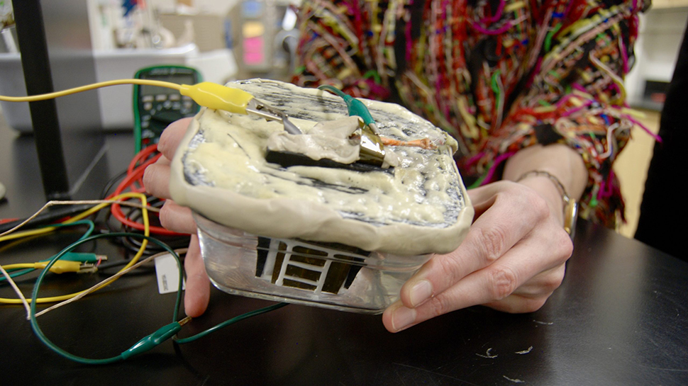
Researchers at the University of Utah’s College of Engineering have pioneered a new form of battery that could help solve this dilemma. The solution, which is at the proof of concept stage, comes in the form of a pyroelectrochemical cell (PEC).
Developed by associate professors of mechanical engineering Roseanne Warren and Shad Roundy, the integrated device harvests ambient thermal energy and converts it into stored electrochemical energy. This effectively creates a supercapacitor or battery, which could be ideal for IoT and sensor applications.
Low levels of energy
The device works by charging with changes in its surrounding temperatures, whether located inside a vehicle, an aircraft, or even underneath soil in an agricultural environment.
“We’re talking very low levels of energy harvesting,” Warren said, “but the ability to have sensors that can be distributed and not need to be recharged in the field is the main advantage. We explored the basic physics of it and found that it could generate a charge with an increase in temperature or a decrease in temperature.”
Whilst solar cells can provide an alternative power source of IoT devices, the practicalities often present issues. “In a lot of environments, you run into two problems,” said Roundy. “One is that it gets dirty over time. Solar cells have to be kept clean. So in these types of applications, they get dirty and their power degrades. And then there are a lot of applications where you just don’t have sunlight available. For example, we work on soil sensors that we put just under the top surface of the soil. You’re not going to get any sunlight.”
With the use of a pyroelectric composite material made of porous polyvinylidene fluoride (PVDF) and barium titanate nanoparticles as the separator in an electrochemical cell, the device’s electrical properties change as it’s heated or cooled. This action modifies the polarization of the pyroelectric separator. This shifting of temperatures in turn creates an electric field within the cell, moving ions around and allowing the cell to store energy.
Sign up to the TechRadar Pro newsletter to get all the top news, opinion, features and guidance your business needs to succeed!
Despite only producing up to 100 microjoules per square centimeter from a single heating/cooling cycle, this could be enough for the needs of some IoT applications.
The study, funded by the National Science Foundation, is the cover feature in the March 21 edition of the journal Energy & Environmental Science, published by the Royal Society of Chemistry.
“Lots of our members call us ‘the magic money tree’,” says Alison Baxter, head of communications for the Authors’ Licensing and Collecting Society (ALCS), based in the United Kingdom. “They don’t really understand where the payments come from,” she says, “but they like getting them.” The ALCS is one of a global group of collecting societies and agencies that compensate authors when their works are copied or shared after publication. This year, the ALCS says, it is due to pay out more than £45 million (US$56 million) and, when money is earned from the use of academic textbooks and research papers, the copyright holders who stand to benefit are often scientists.
ALCS members who claimed for journal or magazine articles this year received around £450 each, on average. Although the sums any individual author is entitled to could be much less than that, the fact remains that researchers who are not members might be missing out on their share.
Collecting societies vary in their exact function, but their common goal is to ensure that authors are remunerated when, for example, a company prints out part of a book to circulate among its staff, or a research paper is printed out and distributed to students. The societies generate income by selling licences that give blanket permission to reproduce copy-righted material, or by gathering payments for the use of specific works.
They then share that money among their members on the basis of which activities generated the funds. Although this is a well-established source of income for many authors and journalists, among researchers there is less awareness of its existence. “My oldest friend is a scientist,” Baxter says, “and it took me a while to convince her that she could claim for her papers by joining the ALCS.”
One reason for a lack of take-up might be cynicism among scientists, and a misapprehension about fraud. “Everyone I have told thinks it’s a scam,” says Nicole Melzack, who is studying for a PhD in energy storage at the University of Southampton, UK, and has been a member of the ALCS since last year. “It’s really hard convincing people that it’s not, but, as long as you own the copyright, which I think most people will for their journal articles, then you have nothing to lose by signing up.”
Careers Collection: Publishing
For those who do, collecting societies can provide a welcome and regular cash flow that requires little or no effort to maintain. Yashar Mousavi, a senior analytical engineer at American Axle & Manufacturing, an automotive engineering firm based in Detroit, Michigan, joined the ALCS as a PhD student at Glasgow Caledonian University, UK, in 2020. “I’ve been paid twice so far, each time between £400 and £600, for papers published in the UK in the journal Chaos, Solitons & Fractals on the topic of fractional calculus and optimization,” he says. “The size of payment depends on many factors, such as the amount of money the ALCS has collected, the number of papers I have shared with them, the percentage of my contribution to the paper, and the journal’s impact factor.”
Even early-career researchers who do not have many publications can benefit, says Melzack. “In 2023, I had one paper published in Frontiers in Energy Research and made £464, and this year I published five papers and got £357,” they say. “It’s great, given the general cost of living and the fact that the academic publishing ecosystem involves so much unpaid labour, so to get something for a paper I’ve written feels validating in some way too.”
Nicole Melzack says that scientists who own the copyright in their publications have nothing to lose by joining a collecting society.Credit: Nicole Melzack
For scientists who publish outside academic journals, the rewards can be even greater. Isabel Thomas, a freelance science writer and children’s book author based in Cambridge, UK, joined the ALCS in 2014. “Since then I’ve had payments every six months, ranging from £77 to £8,000,” she says. “The ALCS also approached a friend of mine who writes practice exam papers and it turned out they were holding almost £30,000 due to her.”
How do collecting societies work?
Collecting societies might seem unusual in the context of academic research, but they are long established in other fields. For example, the Performing Right Society in the United Kingdom and the American Society of Composers, Authors and Publishers in the United States, both founded in 1914, collect fees for music played in public, then distribute the money to the composers and songwriters concerned. Other organizations ensure that artists and photographers are paid when their images are used.
The same principles apply to written work and authors. The ALCS was founded in 1977 by a group of writers who realized that photocopiers were enabling people to reproduce and share works without the creators being compensated. They also set up an accompanying body, the Copyright Licensing Agency (CLA), which collects money that the ALCS then distributes to writers.
The CLA sells and manages collective licences that give organizations the legal right to reproduce copyrighted works (whereas the ALCS handles payments to copyright holders). Baxter says that schools and universities, as well as the UK National Health Service and businesses, all pay the CLA for a licence. “That then means that their staff, students or users are allowed to copy sections of the books they own and share them, both physically and digitally.” The money generated is split between the publishers and authors.
Many other countries have similar collective licensing bodies. The Copyright Agency in Australia, the Indian Reprographic Rights Organisation, CADRA in Argentina and Canada’s Access Copyright all generate revenue through similar processes. The Copyright Clearance Center (CCC) is responsible for similar licences in the United States, but sends payments to publishers for distribution to authors.
Collecting societies also act as advocates and support networks. CADRA, for example, has been particularly successful in attracting researchers, who make up an estimated 40% of its members. Executive director Magdalena Iraizoz, who is based in Buenos Aires, says, “If a scientist has published work, being a member of CADRA not only gives them the benefit of receiving payments for the secondary uses of their works, but also free legal protection against piracy and illegal reproduction.”
How to collect payments
Anyone with publications to their name can join a collecting society and potentially receive payments. Baxter advises that scientists first determine what copyrights they own. “With books, authors aren’t generally asked to sign their copyright away,” she says. “In cases where they do have to, like when publishing in some academic journals, the contract can include a ‘quick clause’ that means the writer can still receive money from us.” These clauses can also apply to work that is published open access.
Authors then need to join the relevant collecting society. This will generally be one based in the country in which their work has been published, although many have reciprocal agreements that allow them to collect income generated overseas that is owed to their members. In its 2022–23 financial year, the Copyright Agency paid out Aus$142 million (US$92 million) to rights holders in Australia and elsewhere. “Most of our direct payments are to Australian writers, artists and publishers,” says a spokesperson for the Copyright Agency. “Most payments from copyright fees we collect for non-Australian works are made via copyright-management organizations similar to us in other countries.”
Science writer Isabel Thomas’s biannual collecting-society payments have ranged from £77 to £8,000.Credit: Elodie Guige
The majority of collecting societies are members of the International Federation of Reproduction Rights Organisations and are listed on its website. Generally, they do not ask individuals for a joining fee and instead take a small percentage from payments they distribute.
Scientists who join the ALCS can register any book with a unique International Standard Book Number (ISBN) product identifier, but only papers or articles published in the past three years in a journal with a UK-based International Standard Serial Number (ISSN) qualify. Baxter recommends that members list anything they think might be eligible. “For journals, we operate on a claim scheme,” she explains. “So, we gather money for a particular ISSN. Then we ask people to tell us what they’ve written and anyone who has contributed to that journal gets a share of the pot.”
CADRA operates in a similar way. “For a scientist to become a member of CADRA, they must have written work published with an Argentinian ISBN or ISSN,” says Iraizoz. “They must then sign the association contract and, once their incorporation is approved, they will be able to be part of the next distribution of rights.” Essentially, researchers list what they own and collecting societies will determine what they’re owed.
A revolutionary new memory device that combines the features of DRAM and NAND flash memory is being developed by a group of researchers at the Korea Advanced Institute of Science and Technology (KAIST).
Led by Professor Shinhyun Choi of the School of Electrical Engineering, the team’s breakthrough promises cheaper, power-efficient solutions that could potentially replace existing memory solutions or be used to implement neuromorphic computing for the next-generation of AI hardware.
According to KAIST, the new device utilizes next-generation phase change memory with ultra-low power consumption, capable of replacing both DRAM and NAND flash memory.
Taking a novel approach
Typically, DRAM offers high-speed performance but is volatile, resulting in data loss when power is shut down. NAND flash memory offers a solution by preserving data even when the power is off, but it does not match the speed of DRAM. This new phase change memory provides a non-volatile, high-speed solution combining the best of both worlds.
Previous versions of phase change memory have had a problem – high power consumption. Despite attempts to reduce consumption by decreasing the physical size of such devices using state-of-the-art lithography technologies, the reductions were minimal, while costs soared.
To overcome this, Professor Choi’s team have established a way to electrically form phase change materials in an extremely small area, successfully developing an ultra-low-power phase change memory device. Notably, this consumes 15 times less power than previous phase change memory models which used expensive lithography tools, a significant breakthrough in the quest for cost and energy efficient memory development.
“The phase change memory device we have developed is significant as it offers a novel approach to solve the lingering problems in producing a memory device at a greatly improved manufacturing cost and energy efficiency, “ said Professor Choi. He went onto to say that he expects this new research to become the basis for future electronic engineering, paving the path for high-density three-dimensional vertical memory and neuromorphic computing systems.
Sign up to the TechRadar Pro newsletter to get all the top news, opinion, features and guidance your business needs to succeed!
This isn’t the only neuromorphic computing solution being worked on at KAIST. Last month scientists there unveiled an AI chip that they claimed can match the speed of Nvidia‘s A100 GPU but with a smaller size and significantly lower power consumption.
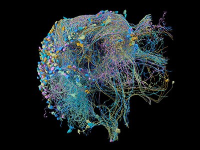
A cubic millimetre is a tiny volume — less than a teardrop. But a cubic millimetre of mouse brain is densely packed with tens of thousands of neurons and other cells in a staggeringly complex architectural weave.
Reconstructing such elaborate arrangements requires monumental effort, but the researchers affiliated with the Machine Intelligence from Cortical Networks (MICrONS) programme pulled it off. It took US$100 million and years of effort by more than 100 scientists, coordinated by 3 groups that had never collaborated before. There were weeks of all-nighters and a painstaking global proofreading effort that continues even now — for a volume that represents just 0.2% of the typical mouse brain. Despite the hurdles, the core of the project — conceived and funded by the US Intelligence Advanced Research Projects Activity (IARPA) — is complete.
Human brain mapping
The resulting package includes a high-resolution 3D electron microscopy reconstruction of the cells and organelles in two separate volumes of the mouse visual cortex, coupled with fluorescent imaging of neuronal activity from the same volumes. Even the coordinators of the MICrONS project, who describe IARPA’s assembly of the consortium as a ‘shotgun wedding’ of parallel research efforts, were pleasantly surprised by the outcome. “It formed this contiguous team, and we’ve been working extremely well together,” says Andreas Tolias, a systems neuroscientist who led the functional imaging effort at Baylor College of Medicine in Houston, Texas. “It’s impressive.”
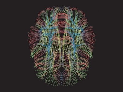
The MICrONS project is a milestone in the field of ‘connectomics’, which aims to unravel the synaptic-scale organization of the brain and chart the circuits that coordinate the organ’s many functions. The data from these first two volumes are already providing the neuroscience community with a valuable resource. But this work is also bringing scientists into strange and challenging new territory. “The main casualty of this information is understanding,” says Jeff Lichtman, a connectomics pioneer at Harvard University in Cambridge, Massachusetts. “The more we know, the harder it is to turn this into a simple, easy-to-understand model of how the brain works.”
Short circuits
There are many ways to look at the brain, but for connectivity researchers, electron microscopy has proved especially powerful.
In 1986, scientists at the University of Cambridge, UK, used serial-section electron microscopy to generate a complete map of the nervous system for the roundworm Caenorhabditiselegans1. That connectome was a landmark achievement in the history of biology. It required the arduous manual annotation and reconstruction of some 8,000 2D images, but yielded a Rosetta Stone for understanding the nervous system of this simple, but important, animal model.
The rise of digital twins
No comparable resource exists for more complex animals, but early forays into the rodent connectome have given hints of what such a map could reveal. Lichtman recalls the assembly he and his colleagues produced in 2015 from a 1,500-cubic-micron section of mouse neocortex — roughly one-millionth of the volume used in the MICrONS project2. “Most people were just shocked to see the density of wires all pushed together in any little part of brain,” he says.
Similarly, Moritz Helmstaedter, a connectomics researcher at the Max Planck Institute for Brain Research in Frankfurt, Germany, says that his team’s efforts3 in reconstructing a densely packed region of the mouse somatosensory cortex, which processes sensations related to touch, in 2019 challenged existing dogma — especially the assumption that neurons in the cortex are randomly wired. “We explicitly proved that wrong,” Helmstaedter says. “We found this extreme precision.” These and other studies have collectively helped to cement the importance of electron-microscopy-based circuit maps as a complement to techniques such as light microscopy and molecular methods.
Bigger and better
IARPA’s motivation for the MICrONS project was grounded in artificial intelligence. The goal was to generate a detailed connectomic map at the cubic-millimetre-scale, which could then be ‘reverse-engineered’ to identify architectural principles that might guide the development of biologically informed artificial neural networks.
Tolias, neuroscientist Sebastian Seung at Princeton University in New Jersey, and neurobiologist Clay Reid at the Allen Institute for Brain Science in Seattle, Washington, had all applied independently for funding to contribute to separate elements of this programme. But IARPA’s programme officers elected to combine the 3 teams into a single consortium — including a broader network of collaborators — issuing $100 million in 2016 to support a 5-year effort.
A Martinotti cell, a small neuron with branching dendrites, with synaptic outputs highlighted.Credit: MICrONS Explorer
The MICrONS team selected two areas from the mouse visual cortex: the aforementioned cubic millimetre, and a much smaller volume that served as a pilot for the workflow. These were chosen so the team could investigate the interactions between disparate regions in the visual pathway, explains Tolias, who oversaw the brain-activity-imaging aspect of the work at Baylor. To achieve that, the researchers genetically engineered a mouse to express a calcium-sensitive ‘reporter gene’, which produces a fluorescent signal whenever a neuron or population of neurons fires. His team then assembled video footage of diverse realistic scenes, which the animal watched with each eye independently for two hours while a microscope tracked neuronal activity.
Probing fine-scale connections in the brain
The mouse was then shipped to Seattle for preparation and imaging of the relevant brain volumes — and the pressure kicked up another notch. Nuno da Costa, a neuroanatomist and associate investigator at the Allen Institute, says he and Tolias compressed their groups’ schedules to accommodate the final, time-consuming stage of digital reconstruction and analysis conducted by Seung’s group. “We really pushed ourselves to deliver — to fail as early as possible so we can course-correct in time,” da Costa says. This meant a race against the clock to excise the tissue, carve it into ultra-thin slices and then image the stained slices with a fleet of 5 electron microscopes. “We invested in this approach where we could buy very old machines, and really automate them to make them super-fast,” says da Costa. The researchers could thus maximize throughput and had backups should a microscope fail.
For phase one of the project, which involved reconstructing the smaller cortical volume, sectioning of the tissue came down to the heroic efforts of Agnes Bodor, a neuroscientist at the Allen Institute, who spent more than a month hand-collecting several thousand 40-nanometre-thick sections of tissue using a diamond-bladed instrument known as a microtome, da Costa says. That manual effort was untenable for the larger volume in phase two of the project, so the Allen team adopted an automated approach. Over 12 days of round-the-clock, supervised work, the team generated almost 28,000 sections containing more than 200,000 cells4. It took six months to image all those sections, yielding some 2 petabytes of data.
The Allen and Baylor teams also collaborated to link the fluorescently imaged cells with their counterparts in the reconstructed connectomic volume.
A network of thousands of individual neurons from a small subset of cells in the Machine Intelligence from Cortical Networks project data set.Credit: MICrONS Explorer
Throughout this process, the Allen team relayed its data sets to the team at Princeton University. Serial-section electron microscopy is a well-established technique, but assembly of the reconstructed volume entails considerable computational work. Images must be precisely aligned with one another while accounting for any preparation- or imaging-associated deformations, and then they are subjected to ‘segmentation’ to identify and annotate neurons, non-neuronal cells such as glia, organelles and other structures. “The revolutionary technology in MICrONS was image alignment,” Seung says. This part is crucial, because a misstep in the positioning of a single slice can derail the remainder of the reconstruction process. Manual curation would be entirely impractical at the cubic-millimetre scale. But through its work in phase one, the team developed a reconstruction workflow that could be scaled up for the larger brain volume, and continuing advances in deep-learning methods made it possible to automate key alignment steps.
To check the work, Sven Dorkenwald, who was a graduate student in Seung’s laboratory and is now a research fellow at the Allen Institute, developed a proofreading framework to refine the team’s reconstructions and ensure their biological fidelity. This approach, which verified the paths of neuronal processes through the connectome, carved the volumes into ‘supervoxels’ — 3D shapes that define segmented cellular or subcellular features, which can be rearranged to improve connectomic accuracy — and Dorkenwald says the final MICrONS data set had 112 billion of them. The system is analogous to the online encyclopedia Wikipedia in some ways, allowing many users to contribute edits in parallel while also logging the history of changes. But even crowdsourced proofreading is slow going — Dorkenwald estimates that each axon (the neuronal projections that transmit signals to other cells) in the MICrONS data set takes up to 50 hours to proofread.
Charting new territory
The MICrONS team published a summary5 of its phase one results in 2022. Much of its other early findings still await publication, including a detailed description of the work from phase two — although this is currently available as a preprint article4. But there are already some important demonstrations of what connectomics at this scale can deliver.
FlyWire: online community for whole-brain connectomics
One MICrONS preprint, for example, describes what is perhaps the most comprehensive circuit map so far for a cortical column6, a layered arrangement of neurons that is thought to be the fundamental organizational unit of the cerebral cortex. The team’s reconstruction yielded a detailed census of all the different cell types residing in the column and revealed previously unknown patterns in how various subtypes of neuron connect with one another. “Inhibitory cells have this remarkable specificity towards some excitatory cell types, even when these excitatory cells are mixed together in the same layer,” says da Costa. Such insights could lead to more precise classification of the cells that boost or suppress circuit activity and reveal the underlying rules that guide the wiring of those circuits.
Crucially, says Tolias, the MICrONS project was about more than the connectome: “It was large-scale, functional imaging of the same mouse.” Much of his team’s work has focused on translating calcium reporter-based activity measurements into next-generation computational models. In 2023, the researchers posted a preprint that describes the creation of a deep-learning-based ‘digital twin’ on the basis of experimentally measured cortical responses to visual stimuli7. The predictions generated by this ‘twin’ can then be tested, further refining the model and enhancing its accuracy.
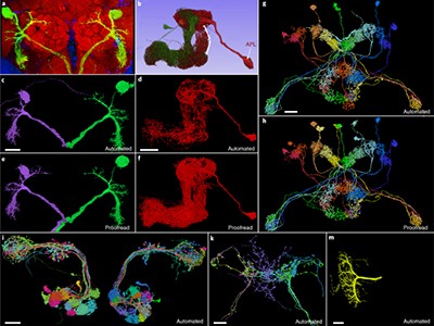
One surprising and valuable product of the MICrONS effort involves fruit flies. Early in the project, Seung’s team began exploring serial-section electron-microscopy data from the Drosophilamelanogaster brain produced by researchers at the Howard Hughes Medical Institute’s Janelia Research Campus in Ashburn, Virginia8. “I realized that because we had developed this image-alignment technology, we had a chance to do something that people thought was impossible,” says Seung. His team — including Dorkenwald — used the Janelia data as a proving ground for the algorithms that had been developed for MICrONS. The result was the first complete assembly of the fruit-fly brain connectome — around 130,000 neurons in total9.
Given that the wiring of the nervous system is generally conserved across fruit flies, Dorkenwald is enthusiastic about how these data — which are publicly accessible at http://flywire.ai — could enable future experiments. “You can do functional imaging on a fly, and because you can find the same neurons over in the connectome, you will be able to do these functional-structure analyses,” he says.
The mouse connectome will not be so simple, because connectivity varies from individual to individual. But the MICrONS data are nevertheless valuable for the neuroscience community, says Helmstaedter, who was not part of the MICrONS project. “It’s great data, and it’s inspiring people just to go look at it and see it,” he says. There’s also the power of demonstrating what is possible, and how it could be done better. “You’ve got to do something brute force first to find out where you can make it easier the next round,” says Kristen Harris, a neuroscientist at the University of Texas at Austin. “And the act of doing it — just getting the job done — is just spectacular.”
Terra incognita
Even as analysis of the MICrONS data set proceeds, its limitations are already becoming clear. For one thing, volumes from other distinct cortical regions will be needed to identify features that are broadly observed throughout the brain versus those features that are distinct to the visual cortex. And many axons from this first cubic millimetre will inevitably connect to points unknown, Lichtman notes, limiting researchers’ ability to fully understand the structure and function of the circuits within it.
Scaling up will be even harder. Lichtman estimates that a whole-brain electron-microscopy reconstruction would produce roughly an exabyte of data, which is equivalent to a billion gigabytes and is 1,000 times greater than the petabytes of data produced by the MICrONS project. “This may be a ‘Mars shot’ — it’s really much harder than going to the Moon,” he says.
Still, the race is under way. One major effort is BRAIN CONNECTS, a project backed by the US National Institutes of Health with $150 million in funding, which is coordinated by multiple researchers, including Seung, da Costa and Lichtman. “We’re not delivering the whole mouse brain yet, but testing if it’s possible,” da Costa says. “Mitigating all the risks, bringing the cost down, and seeing if we can actually prepare a whole-mouse-brain or whole-hemisphere sample.”
In parallel, Lichtman is working with a team at Google Research in Mountain View, California, led by computer scientist Viren Jain — who collaborated with MICrONS and is also part of the BRAIN CONNECTS leadership team — to map sizable volumes of the human cortex using electron microscopy. They’ve already released data from their first cubic millimetre and have plans to begin charting other regions from people with various neurological conditions10.
NatureTech hub
These efforts will require improved tools. The serial-section electron-microscopy strategy that MICrONS used is too labour-intensive to use at larger scales and yields relatively low-quality data that are hard to analyse. But alternatives are emerging. For example, ‘block-face’ electron-microscopy methods, in which the sample is imaged as a solid volume and then gradually shaved away with a high-intensity ion-beam, require less work in terms of image alignment and can be applied to thick sections of tissue that are easier to manage. These methods can be combined with cutting-edge multi-beam scanning electron microscopes, which image specimens using up to 91 electron beams simultaneously, thus accelerating data collection. “That’s one of the leading contenders for scale up to a whole mouse brain,” says Seung, who will be working with Lichtman on this strategy.
Further automation and more artificial-intelligence tools will also be assets. Helmstaedter and his colleagues have been looking into ways to simplify image assembly with an automated segmentation algorithm called RoboEM, which traces neural processes with minimal human intervention and can potentially eliminate a lot of the current proofreading burden11. Still, higher-quality sample preparation and imaging are probably the true key to efficiency at scale, Helmstaedter says. “The better your data, the less you have to worry about automation.”
However they are generated, making sense of these connectome maps will take more than fancy technology. Tolias thinks “it will be almost impossible” to replicate the coupling of structure and activity produced by MICrONS at the whole-brain scale. But it’s also unclear whether that will be necessary and to what extent functional information can be inferred through a better understanding of brain structure and organization.
For Lichtman, the connectome’s value will ultimately transcend conventional hypothesis-driven science. A connectome “forces you to see things you weren’t looking for, and yet they’re staring you in the face”, he says. “I think if we do a whole mouse brain, there will be just an infinite number of ‘wow, really?’ discoveries.”
Losing a funding competition didn’t set Ellen Stofan back — instead, she did a career pivot, and came across new opportunities.Credit: NASA/Joel Kowsky
In 2021, planetary scientist Ellen Stofan was appointed undersecretary of science and research at the Smithsonian Institution in Washington DC, the US national research and museum complex. There, she oversees its scientific research centres as well as the National Air and Space Museum, the National Museum of Natural History and the National Zoo and Conservation Biology Institute. Before this, she was director of the Smithsonian’s National Air and Space Museum, where she launched a 7-year restoration of the building and oversaw celebrations marking 50 years since the first Moon landing. Stofan’s doctoral research at Brown University in Providence, Rhode Island, focused on the geology of Venus.
Before joining the Smithsonian, she spent some 25 years working in space-related organizations — including NASA’s Jet Propulsion Laboratory and as the agency’s chief scientist. She helped to develop NASA’s plan to get humans to Mars and worked on the Magellan mission to Venus and the 13-year Cassini mission that documented Saturn and its moons.
Describe a typical day.
My portfolio is really broad, so there’s no typical day. I might be having a meeting about bringing pandas back to the zoo in Washington DC, or discussing how to dispose of the Smithsonian’s collection of human remains in an ethical way. Or talking about the budget — it’s always the budget.
Training: Persuasive grant writing
Is discussing the budget what you thought you would be doing at the start of your career?
Probably not, but the budget reflects the organization’s strategy and priorities, so you have to understand why you are putting money in certain areas. Speaking of priorities, over the past few years, I’ve been working on the Our Shared Future: Life on a Sustainable Planet research initiative, which we announced at the United Nations climate conference COP 27 two years ago. What’s amazing is the amount of science we were already doing along those lines. For example, in Montana, we have been recreating the ecosystem of an American prairie — we’ve reintroduced bison, and all of a sudden birds and insects have started coming back.
Did you plan to work in the museum sector?
I interned at the Air and Space Museum when I was an undergraduate, but at that time I just wanted to be a geologist, write papers and maybe work at a university. A thread through my career is working in great teams — that was why I enjoyed NASA so much. To explore Venus or the moons of Saturn, you have to put together an engaged team by bringing together people with different skills and ideas. At NASA, I led a team that was bidding for a Discovery Program grant, which can be used to fund smaller planetary missions using fewer resources and with shorter development times. Our proposed mission, the Titan Mare Explorer vessel, would explore the seas of liquid hydrocarbons, such as methane and ethane, on Titan, Saturn’s largest moon. Working with the fun, smart, creative and innovative people on the team did not feel like work at all. Our project was one of the three finalists in 2012, but another one was chosen.
How did that feel?
Not getting the grant was devastating — not just for me, but for the team. I felt like I had let them down. For a while, I couldn’t talk about the project without crying. I thought about leaving science, because I didn’t see how anything could ever match that.
It took me months to process it all. Before our bid, NASA had concluded that no research projects could reach the outer Solar System for less than a billion dollars. We were bidding for around US$400 million, and our proposal helped to pioneer the idea that, through innovation and judicious use of technology, these projects could be done more cheaply. Our mission created this small paradigm shift — and, all of a sudden, we saw people proposing projects that would go to the outer Solar System at much lower costs than before.
The display of Amelia Earhart’s plane at the Smithsonian’s National Air and Space Museum.Credit: Jacquelyn Martin/AP Photo/Alamy
What is your approach to career setbacks?
You want to be the kind of person who shrugs off failure — but it’s hard. Everyone goes through it. When I was still processing losing the grant, I was invited to interview to be chief scientist of NASA. I got the job and held that position for three years. My career went a whole different way — I left NASA in 2016, and then the Smithsonian job came up.
Is the Titan Mare project still ongoing?
No, but I’m a co-investigator on a mission called Dragonfly. This drone will launch in late 2026 and will land on Titan in the 2030s. It’s going to fly around the equatorial region, where we think standing pools of liquid methane and liquid ethane might exist. There’s a lot of debate in the scientific community right now about whether life could ever exist on a body like Titan. What we will be able to learn about ‘prebiotic chemistry’ — the study of how chemical compounds assembled to form the precursors to life — from the mission is really exciting.
Did you always dream of a career in space exploration?
Not when I was younger, because my father was an engineer at NASA and the only people he worked with were men — so I just didn’t think it was a place for me. It was only by reading in National Geographic about primatologist and anthropologist Jane Goodall and palaeoanthropoligst Mary Leakey, who studied human origins in Africa, that I realized that not only could women do science, but they could be famous scientists.
When I began my career in the 1980s, I was often either the only woman in the room, or one of the few. And some people thought that I didn’t belong in the room, because I was a woman. I had enough confidence to think, “What’s your problem?”
Things have changed a lot, but women are still under-represented in physics, engineering and computer science, and we’re not tapping into the talent. Hiring people from groups that are under-represented in science is not about achieving diversity for diversity’s sake. We know from scientific research that diverse teams perform better.
At NASA, I looked at our workforce and thought about whether we were tapping into the best talent. People often talk about diversity, but they forget about inclusion. NASA was sensitive to this after the Challenger accident — the space shuttle broke apart seconds after take off in 1986, killing all seven members of the crew. One of the findings was that managers were not listening to their teams. It’s important to create an environment in which everyone can contribute and participate. Even if you have a diverse workforce, if you don’t make people feel included, they’re not going to stay.
What is a key priority for you at the Smithsonian?
When we were redoing the museum, one important part of our mission was to inspire the next generation of innovators and explorers. Are we telling stories so that every kid who comes into the museum, no matter their race, gender or other aspect of their life, is going to find someone who looks like them?
In the past, the story of space centered charismatic figures, such as astronaut Neil Armstrong — but look at the success of the 2016 movie HiddenFigures, which is about a team of Black female mathematicians working for NASA during its early years. Visitors might notice that, at the museum, we’re telling a much broader range of stories. In February, the first private company, in partnership with NASA, touched down on the Moon; there are now many more countries involved in space exploration, and private individuals are going into space. The story of space is changing.
Do you have a favourite museum exhibit?
We have an X-wing fighter from the Star Wars films, which I absolutely love. We’ve also had the Starship Enterprise from the Star Trek series.
But my absolute favourite is aviator Amelia Earhart’s Lockheed Vega aeroplane. It’s this cheeky red colour that, to me, symbolizes her saying, ‘I’m going to fly despite what anyone thinks.’
Would you ever like to go into space?
When I went to my first launch, the rocket blew up. It was uncrewed, but it’s seared into my memory. I’m not terribly adventurous. I’m happy to be an armchair explorer.