Categories
Life Style

Exploring the lung microbiome’s role in disease

[ad_1]

Conceptual depiction of lungs containing microbes.

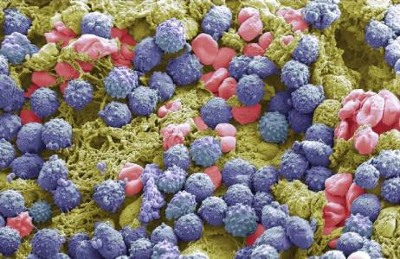
Credit: Stephan Schmitz/Folio Art

Not so long ago, the textbook image of the lungs was that of a sterile environment. “When I was in medical school, around 2005, literally my pathology textbook said that the normal lung is free from bacteria,” recalls Robert Dickson, a pulmonary and critical-care physician at the University of Michigan in Ann Arbor. “This was dogma for more than a century.” But over the past decade, that picture has gradually been scrubbed away as sampling of the lungs has unmasked a community of microorganisms hidden inside — albeit an unusual one.

The lung menagerie looks nothing like the microbial rainforest that thrives in the fertile gut; by comparison, the lungs are a veritable desert. “The quantity is really low — many orders of magnitude lower than the upper respiratory tract, never mind the gastrointestinal tract,” says Ronald Collman, a microbiologist and pulmonary physician at the University of Pennsylvania in Philadelphia.

The lungs’ microbial community is also notably more transient than that of the gut. The body has evolved ways to keep the lungs clean, so in healthy lungs there are few or no resident replicating bacteria. Instead, the lungs host a constant flux of microbes that mostly mirror the diverse community of the upper airways, especially around the back of the throat and the vocal cords.

But the systems that prevent the warm, wet lungs from being the perfect accommodation for bacteria can degrade. In people with chronic conditions, Dickson says, lung tissue becomes inflamed and the environment changes — mucus production increases, airway tissue swells, nutrients become more readily available to bacteria and potentially damaging strains such as Pseudomonas and Haemophilus influenzae can bloom and become resident.

Blue bacteria pictured on a brown background

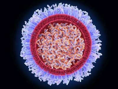
Pseudomonas bacteria can cause lung infections.Credit: David M. Phillips/SPL

Most strikingly, there are signs that a shift in the lung microbiota might begin in advance of some conditions, such as chronic obstructive pulmonary disease (COPD) and lung cancer, and support their development. If proved correct, this could make lung microbes a target for intervention to prevent or delay disease. “One might be able to do respiratory microbiome therapeutics,” says Collman.

A healthy state of flux

Although it is now accepted that a healthy lung is not sterile, researchers have not yet been able to define what the microbial contents of a normal lung should be. “We don’t know yet how to best define a healthy microbiome,” says Yvonne Huang, a lung-disease specialist at the University of Michigan. She recalls that she spent two days as part of a specialist committee for the US National Academy of Sciences debating and failing to agree on such a definition in 2017.

Some bacteria do seem to be common in the lungs of healthy people. “Certain microbes keep coming up,” says Stavros Garantziotis, a lung researcher at the US National Institute of Environmental Health Sciences in Durham, North Carolina. Key players include Prevotella, Streptococcus and Veillonella species, all bacterial residents of the upper airways. They probably enter the lungs through the inhalation of small droplets while people are sleeping — something that occurred in around half of a group of healthy adults in a study involving a radioactive tracer, to varying extents1.

Under typical circumstances, even these bacteria are more like regular tourists to the lung than residents — the body continually works to remove them. “Think about this dynamic community as like a train station, with people coming and going, over and over,” says Leopoldo Segal, a lung clinician at NYU Langone Health in New York City. How many microbes are visiting the lungs at a given time seems to vary from person to person and throughout an individual’s life. In a study of 49 adults with healthy lungs, Segal and his colleagues found that almost half had a relatively high load of oral microbes in their lungs2. They also found that those with high bacterial load had more infection-fighting white blood cells and pro-inflammatory molecules in their lungs.

Person in white coat and gloves holding pipette, standing by laboratory bench

Yvonne Huang, a lung-disease specialist at the University of Michigan in Ann Arbor, says that researchers do not yet know how best to define a healthy lung microbiome.Credit: Guowu Bian

Some evidence suggests that this immune activity could be beneficial to health. In a 2021 study, human oral bacteria were infused into the lower airways of mice, causing dysfunctional changes in the distribution of microbiota in the lungs, known as dysbiosis. This was rapidly cleared by the mice, but caused a prolonged immune response that made them less susceptible to Streptococcus pneumoniae, a cause of pneumonia3. “Benign commensals may have some beneficial roles in priming your immune system to respond better to a pathogen,” says Segal. However, aspiration of oral bacteria could also exacerbate inflammatory injury, he adds.

“There is a lung microbiome that seems to contribute to health,” says Garantziotis. Conversely, some bacteria seem to be associated with lung disease. “It is probably logical to assume certain bacteria in your lungs will predispose you to inflammation,” he says.

Disease links

Across dozens of studies and hundreds of volunteers, much the same assortment of bacteria turns up in the lungs of people with chronic conditions such as COPD and the scarring and thickening of lung tissue known as pulmonary fibrosis. The changes in quantity or type of lung microbiota are typically subtle, and not enough to be called an infection. “There’s a host of diseases where we see disruption of the normal lung microbiome, but it’s not dramatic like in infections,” says Collman. Even so, researchers are keen to know what involvement lung microbiota might have in disease. “We’re asking if a disordered microbiota is driving a dysregulated immune response that’s contributing to injury,” says Dickson.

Person wearing suit and tie standing to right of image with arms folded

Robert Dickson is a pulmonary and critical-care physician at the University of Michigan in Ann Arbor.Credit: Michigan Photography

Some evidence does link bacterial load in the lungs to health outcomes. “The more bacteria we find in the lungs, the worse patients do,” says Dickson. A study of more than 300 people on mechanical ventilation linked the presence of more Staphylococcus and Pseudomonas strains in the lower airways with greater inflammation and reduced survival after 30 days4. Outcomes for people who have received lung transplants also correlate with bacterial load5. “The quantity of bacteria DNA we find in the lungs of transplant patients predicts who’s going to experience rejection and ultimately die,” Dickson says.

Although the connections between lung microbiota and health are apparent, the directionality is not — do microbes in the lungs contribute to disease, or are they simply opportunist squatters taking advantage of the disease state? Certainly, disease can make the lungs more hospitable to microbe entry or replication. “Distinguishing cause from effect is really tough in human studies of COPD, because you see destruction of tissue, excess mucus production — things that may encourage bacteria overgrowth,” says Collman.

A precise timeline is arduous to follow in individuals, especially because the gold-standard procedure for sampling the lung microbiota — a bronchoscopy — is invasive and unpleasant, and therefore undertaken only when there is a compelling benefit. “The lungs are hard to sample,” says Michael Cox, a respiratory microbiome researcher at the University of Birmingham, UK. “You must go through the mouth,” he explains, which also makes contamination with oral microbiota difficult to avoid.

In 2016, a study in long-tailed macaques (Macaca fascicularis) became the first to examine the dynamics of the lung microbiota over time in primates6. The team, led by researchers at the University of Pittsburgh in Pennsylvania, monitored the lungs of macaques that had an HIV-like immunosuppressive infection. They found that oral bacteria progressively accumulated in the lungs, and that the animals subsequently developed COPD-like changes. “These findings suggest that changes in the lung microbiome might contribute to the development of COPD,” says Alison Morris, a pulmonary specialist at the University of Pittsburgh who worked on the study.

The true nature of the relationship between the lung microbiota and chronic lung disease probably lies somewhere in between. “I don’t think it’s a unidirectional cause and effect,” says Huang, who has studied COPD and asthma. A deteriorating lung allows more microbes to gain entry and impairs clearance mechanisms, but greater commuting of bacteria from the upper respiratory tract could also elicit a greater response from immune cells and ramp-up inflammation. “Particular culprits might be playing a part in helping move forward the inflammatory process, leading to further inflammation and damage,” Huang says.

Beyond the lungs

Emerging connections between lung microbiota and disease are not limited to COPD — or even to the lungs at all. A connection between immune cells in the lung and diseases of the brain is also coming into view. In 2022, researchers at the University of Göttingen in Germany revealed that changes to rats’ lung microbiota influenced the animals’ susceptibility to developing multiple sclerosis (MS), an autoimmune disease of the central nervous system7. It seemed that certain immune cells in the brain, known as microglia, were influenced by microbial signalling in the lungs — behaviour that the researchers described as a “remote warning system” for the brain. This insight has led some people in the field to consider whether inhaled probiotics could one day be deployed as a treatment for MS.

Evidence of a link between lung microbes and cancer is also growing. It is already widely accepted that certain gut microbes can directly increase cancer risks, a notable example being Helicobacter pylori and stomach cancer8. There are similar concerns about the Mycobacterium tuberculosis bacteria that infect the lungs and cause tuberculosis (TB). In 2009, a review of 41 studies found that the risk of developing lung cancer was significantly higher in people with a history of TB9. More than a decade later, in 2021, a study of around 20,000 people in Taiwan concluded that cancer of the gut, breast, kidney and thyroid was more likely to spread to the lungs in people who were infected with M. tuberculosis10. Researchers suggest that metabolites from bacteria could damage the DNA of lung tissue or stoke lung inflammation, creating fertile ground for tumours.

Close-up of bacteria coloured purple and yellow

Mycobacterium tuberculosis bacteria cause the lung infection tuberculosis and have been linked to lung cancer.Credit: A. Dowsett, National Infection Service/SPL

Around the same time, Segal and his colleagues presented a study of 83 people with lung cancer in New York City that, for the first time, demonstrated that dysbiosis in the lungs affects lung tumour progression and clinical prognosis, as shown by decreased survival among people with dysbiosis and early-stage disease11. The bacteria most associated with this dysbiosis was Veillonella parvula, a microbe commonly found in the mouth, but one that is sometimes linked to infections such as gum disease. Segal and his colleagues seeded the lungs of mice with V. parvula. In mice with lung cancer, exposure to the microbe decreased survival and stoked up inflammatory markers such as IL-17. “Dysbiosis turns on an inflammatory cascade that fuels tumour progression,” says Segal. Blocking the IL-17 pathway reduced the effect of dysbiosis on tumour progression, suggesting a potential path to slowing down lung cancer.

Therapeutic aspirations

As knowledge of the lung microbiota improves, medical researchers are turning their attention to potential practical applications, such as intervening to maintain or restore lung health.

One approach to dealing with dysbiosis could be to get rid of unwanted microbes using targeted antibiotics — either in the lungs directly, or in the upper airways. “You might want to target the upper airway microbiome in disease, because that is the source of some of these microbes that reach the lungs,” says Segal. This could be especially beneficial to people who happen to aspirate more bacteria into their lungs than is considered typical, and who are therefore vulnerable to dysbiosis.

However, the effect of such an intervention can be unpredictable. In one trial that gave erythromycin — an antibiotic used to treat inflammatory airway disease — to people with lung damage, it was found that in some individuals the drug served only to replace one lot of microbes with a strain of Pseudomonas aeruginosa that was more resistant to antibiotics12.

Another strategy could be to airdrop a healthier microbial community into the lower airways, to displace unwanted residents. “The notion of a therapeutic respiratory tract repopulation is, I think, plausible,” says Collman. Which strains and in what proportions is unknown, however. “I don’t think we are quite at the mechanistic understanding for the next phase of trying to develop therapeutic approaches,” Collman says.

Some researchers are looking at lung microbiota in a different way — not as a target for therapy itself, but as a source of information that could guide the selection of existing therapies. For example, Huang’s group has found differences in the airway or lung microbiota of people who do and do not respond to inhaled steroids — drugs that are commonly prescribed for people with COPD and asthma13. She wants to further investigate what nuances in the microbiota might affect how well people respond to treatments.

“Ultimately, we need randomized controlled trials, where we ask if variation in the microbiome explains differential benefits of therapies,” says Dickson. Trials of this nature could not only help to steer treatment decisions, but also help to answer the question of directionality that plagues the studies of the links between the lung microbiota and disease. “If we can show that differential treatments work or don’t work based on your lung microbiota,” Dickson says, “that would be the most compelling argument for causality.”

[ad_2]

Source Article Link

Categories
Life Style

Diabetes drug slows development of Parkinson’s disease

[ad_1]

A diabetes drug related to the latest generation of obesity drugs can slow the development of the symptoms of Parkinson’s disease, a clinical trial suggests1. Participants who took the drug, called lixisenatide, for 12 months showed no worsening of their symptoms — a gain in a condition marked by progressive loss of motor control.

Further work is needed to control side effects and determine the best dose, but researchers say that the trial marks another promising step in the decades-long effort to tackle the common and debilitating disorder.

“This is the first large-scale, multicentre clinical trial to provide the signs of efficacy that have been sought for so many years,” says Olivier Rascol, a Parkinson’s researcher at Toulouse University Hospital in France, who led the study.

The diabetes connection

Lixisenatide is a glucagon-like peptide-1 (GLP-1) receptor agonist, making it part of a large family of similar compounds used to treat diabetes and, more recently, obesity. (The weight-loss drug semaglutide, sold under the brand name Wegovy, is a GLP-1 compound.)

Many studies have shown a link between diabetes and Parkinson’s2. People with diabetes are around 40% more likely to develop Parkinson’s. And people who have both Parkinson’s and diabetes often see more rapid progression of symptoms than do those who have only Parkinson’s.

Animal studies3 have suggested that some GLP-1 drugs, which influence levels of insulin and glucose, can slow the symptoms of Parkinson’s. Smaller trials, published in 20134 and 20175, suggested that the GLP-1 molecule exenatide, another diabetes drug, could do the same in people.

Progression halted

In the latest, larger study, the French researchers investigated lixisenatide in 156 people with mild to moderate Parkinson’s symptoms, all of whom were already taking the standard Parkinson’s drug levodopa or other drugs. Half got the GLP-1 drug for a year and the others received a placebo.

After 12 months, those in the control group showed a worsening of their symptoms. Specifically, their score had increased by three points on a scale used to assess the severity of Parkinson’s that measures how well people can perform tasks including speaking, eating and walking.

Those taking the drug had no change in their scores on this scale. But the treatment did induce side effects. Nausea occurred in nearly half, and vomiting in 13%, of people on the medication. The results are published in The New England Journal of Medicine.

Not a miracle drug

David Standaert, a neurologist at the University of Alabama at Birmingham, who was not involved in the trial, says it’s important to know whether the effect will last beyond a year.

“We’re all cautious. There’s a long history of trying different things in Parkinson’s that ultimately didn’t work,” he says. A difference of three points in the rating score is a small change — one that many people with Parkinson’s would struggle to notice, he says. “What happens at 5 years? Is it 15 points then, or is it still 3? If it’s still 3, then this is not worth it.”

Lixisenatide as a diabetes treatment was pulled from the US market last year by its Paris-based manufacturer Sanofi for commercial reasons. But Standaert says that this would not have affected development of a possible treatment for Parkinson’s, because other GLP-1 drugs are available.

“I view this as a study of the class. I don’t know if this particular one is the right answer,” he says. Newer GLP-1 drugs (lixisenatide was developed in the 2000s) could offer fewer and milder side effects or work at lower doses, he adds.

Another question that needs further consideration is just how some GLP-1 drugs might protect against Parkinson’s. The compounds are known to reduce inflammation, which has led some researchers to suggest that they prevent the steady loss of dopamine-producing neurons that drives the condition. That would offer a significant benefit over existing treatments such as levodopa, which mask the symptoms but don’t address the underlying cause. But this trial and others haven’t assessed neuron loss.

Researchers are now waiting for the results of a large clinical trial examining the effects of a two-year course of exenatide in people with Parkinson’s disease. Those data will be available in the second half of this year, according to Tom Foltynie, a neurologist at University College London, in comments provided to the UK Science Media Centre.

[ad_2]

Source Article Link

Categories
Life Style

mRNA drug offers hope for treating a devastating childhood disease

[ad_1]

A drug that uses messenger RNA technology has shown early success in addressing the core deficiency behind a rare genetic disorder. The results have ignited hope that the technology — which first gained attention through its breakthrough use in COVID-19 vaccines — could realize its long-awaited promise of generating therapeutic proteins directly in the body.

This clinical advance, reported today in Nature1, provides a boost to current mRNA applications, which remain limited to vaccines.

“This is a first step in the right direction,” says Katalin Karikó, a Nobel prizewinning pioneer of mRNA technologies who is affiliated with the University of Szeged in Hungary and the University of Pennsylvania in Philadelphia.

Yet challenges remain — especially the fleeting nature of mRNA and the side effects it causes, which complicate the path towards widespread adoption.

Metabolic makeover

Designed by Moderna in Cambridge, Massachusetts, the current therapy uses mRNA technology to restore metabolic function in people with propionic acidaemia.

This rare genetic disorder, which affects about one in 100,000 individuals worldwide, arises from mutations in either of two genes that together encode an enzyme necessary for the efficient breakdown of certain protein components. Without this enzyme, cells can’t process some nutrients properly.

That leads to the accumulation of toxic chemicals in the blood and tissues, and damages vital organs, including the heart and the brain. Symptoms, such as vomiting, usually start within the first few days after birth.

People can manage the condition with measures such as special diets. But there are currently no treatments that tackle the underlying cause directly.

Moderna’s drug, known as mRNA-3927, aims to address that gap. It contains two mRNA sequences that each craft parts of the otherwise faulty enzyme. These mRNAs are encased in a tiny fat bubble — called a lipid nanoparticle — similar to the carrier used in the company’s COVID-19 vaccine.

The therapeutic mRNA drug is administered slowly through hours-long infusions every two or three weeks. It is also given in doses hundreds of times greater than those of COVID-19 vaccines. Once the therapy enters the bloodstream, the lipid nanoparticles help to direct the mRNA to cells in the liver, where the functional enzyme is made.

Trade-offs and benefits

Initial results from a small trial of mRNA-3927 indicate that the restoration of enzymatic activity is beneficial. Eight of the 16 participants had experienced life-threatening episodes connected to their impaired metabolism in the year before starting treatment. For those eight, the likelihood of experiencing another such event decreased by an average of 70–80% while taking the therapy.

This outcome, based on a small number of people, did not reach the threshold of statistical significance. Nonetheless, “it’s a very encouraging step”, says Jerry Vockley, a medical geneticist at the University of Pittsburgh Medical Center in Pennsylvania who helped to design the trial but who was not involved in its execution.

According to Kyle Holen, head of therapeutics development at Moderna, the company is now recruiting more trial participants as it advances mRNA-3927 towards the goal of marketing approval.

Moderna is also analysing other outcome measures related to quality-of-life metrics — indicators that, anecdotally at least, seem to be improving for some recipients of the treatment.

Nassrine Fawaz in Livonia, Michigan, has witnessed a transformation in her 4-year-old daughter, who has received mRNA-3927 for the past 2.5 years. After each infusion, “she’s focused, she’s energetic, she’s up and ready for the day — all of those great things”.

Room for improvement

Developers of mRNA therapeutics had long worried that repeated administration might trigger immune responses against the treatment. However, with individuals having now received regular infusions of mRNA for months or even years without issue, this concern has been alleviated.

“That’s pretty big,” says Alex Wesselhoeft, director of RNA therapeutics at Mass General Brigham’s Gene and Cell Therapy Institute in Cambridge, Massachusetts.

But there are trade-offs: most people reported side effects in response to the treatment. These ranged from infections to severe swelling of the pancreas. However, as study investigator Andreas Schulze, a metabolic-disease specialist at the Hospital for Sick Children in Toronto, Canada, points out, many of the reactions are more likely to be “related to the underlying disease” than to the treatment.

Still, with a side-effect profile close to what Wesselhoeft describes as the “upper limit of tolerability”, and only modest clinical gains, he and others think that further refinements are needed before mRNA technologies can provide a fully corrective and long-term solution to genetic diseases.

“I’m just doubtful this is going to be a long-term therapy,” says Romesh Subramanian, a biotechnology consultant in Framingham, Massachusetts, who, in a previous job, worked in collaboration with Moderna scientists to develop mRNA therapies for rare diseases. “I think it needs to be much less frequent dosing with better [nanoparticles] or more potent mRNA.”

Meanwhile, many families affected by propionic acidaemia are maintaining a wait-and-see attitude. “The verdict is still out,” says Jill Chertow, founder and president of the Propionic Acidemia Foundation, a non-profit organization based in Deerfield, Illinois.

“We can only be hopeful since, right now, that’s all that we have.”

[ad_2]

Source Article Link

Categories
Life Style

Time to sound the alarm about the hidden epidemic of kidney disease

[ad_1]

Coloured 3D computed tomography scan of healthy human kidneys.

Kidney disease is growing worldwide. The secretariat of the World Health Organization has welcomed the call to include it as a non-communicable disease that causes premature deaths.Credit: Vsevolod Zviryk/SPL

A quiet epidemic is building around the world. It is the third-fastest-growing cause of death globally. By 2040, it is expected to become the fifth-highest cause of years of life lost. Already, 850 million people are affected, and treating them is draining public-health coffers: the US government-funded health-care plan Medicare alone spends US$130 billion to do so each year. The culprit is kidney disease, a condition in which damage to the kidneys prevents them from filtering the blood.

And yet, in discussions of priorities for global public health, the words ‘kidney disease’ do not always feature. One reason for this is that kidney disease is not on the World Health Organization (WHO) list of priority non-communicable diseases (NCDs) that cause premature deaths. The roster of such NCDs includes heart disease, stroke, diabetes, cancer and chronic lung disease. With kidney disease missing, awareness of its growing impact remains low.

The authors of an article in Nature Reviews Nephrology this week want to change that (A. Francis et al. Nature Rev. Nephrol. https://doi.org/10.1038/s41581-024-00820-6; 2024). They are led by the three largest professional organizations working in kidney health — the International Society of Nephrology, the American Society of Nephrology and the European Renal Association — and they’re urging the WHO to include kidney disease on the priority NCD list.

This will, the authors argue, bring attention to the growing threat, which is particularly dire for people in low- and lower-middle-income countries, who already bear two‑thirds of the world’s kidney-disease burden. Adding kidney disease to the list will also mean that reducing deaths from it could become more of a priority for the United Nations Sustainable Development Goals target to reduce premature deaths from NCDs by one-third by 2030.

As of now, rates of chronic kidney disease are likely to increase in low- and lower-middle-income countries as the proportion of older people in their populations increases. Inclusion on the WHO list could provide an incentive for health authorities to prioritize treatments, data collection and other research, along with funding, as with other NCDs.

Kidney disease often accompanies other conditions that do appear on the NCD list, such as heart disease, cancer and diabetes — indeed, kidney-disease deaths caused specifically by diabetes are on the list. But the article authors argue that “tackling diabetes and heart disease alone will not target the core drivers of a large proportion of kidney diseases”. Both acute and chronic kidney disease can have many causes. They can be caused by infection or exposure to toxic substances. Increasingly, the consequences of global climate change, including high temperatures and reduced availability of fresh water, are thought to be contributing to the global burden of kidney disease, as well.

Light micrograph of the kidney glomerulus

The kidney glomerulus filters waste products from the blood. In people with damaged kidneys, this happens through dialysis.Credit: Ziad M. El-Zaatari/SPL

The WHO secretariat, which works closely with the nephrology community, welcomes the call to include kidney disease as an NCD that causes premature deaths, says Slim Slama, who heads the NCD unit at the secretariat in Geneva, Switzerland. The data support including kidney disease as an NCD driver of premature death, he adds.

The decision to include kidney disease along with other priority NCDs isn’t only down to the WHO, however. There must be conversations between the secretariat, WHO member states, the nephrology community, patient advocates and others. WHO member states need to instruct the agency to take the steps to make it happen, including providing appropriate funding for strategic and technical assistance.

Data and funding gaps

Three reports based on surveys by the International Society of Nephrology since 2016 highlight the scale of data gaps (A. K. Bello et al. Lancet Glob. Health 12, E382–E395; 2024). In many countries, screening for kidney disease is difficult to access and a large proportion of cases go undetected and therefore uncounted. For example, it is not known precisely how many people with kidney failure die each year because of lack of access to dialysis or transplantation: the numbers are somewhere between two million and seven million, according to the WHO. Advocates must push public-health officials in more countries to collect the data needed to monitor kidney disease and the impact of prevention and treatment efforts.

Even with better data, treatments for kidney disease are often prohibitively expensive. They include dialysis, an intervention to filter the blood when kidneys cannot. Dialysis is often required two or three times weekly for the remainder of the recipient’s life, or until they can receive a transplant, and it is notoriously costly. In Thailand, for example, it accounted for 3% of the country’s total health-care expenditures in 2022, according to the country’s parliamentary budget office.

These costs could come down if people who have diabetes or high blood pressure, for example, could be routinely screened for impaired kidney function, because they are at high risk of developing chronic kidney disease. This would enable kidney damage to be detected early, before symptoms set in, opening the way for treatments that do not immediately require dialysis or transplant surgery.

New drugs that boost weight loss and treat type 2 diabetes could also help to prevent or reduce stress on the kidneys, but these, too, are too expensive for many people in need. That is why something needs to be done to make drugs more affordable. The pharmaceutical industry, which has become extremely profitable, has a crucial role. In Denmark, for example, the industry’s profits helped to tip the national economy from recession into growth in 2023, according to the public agency Statistics Denmark. The COVID-19 pandemic showed that making profits and making drugs available, and affordable, to a wide population need not be mutually exclusive. Similarly innovative thinking is now needed. “The whole world needs to reckon with this kidney problem,” says Valerie Luyckx, a biomedical ethicist at the University of Zurich in Switzerland.

The WHO adding kidney disease to its priority list could also attract funding for treatment, research and disease registries. That could jump-start the development of new treatments and help to make current treatments more affordable and accessible.

NCDs are responsible for 74% of deaths worldwide, but the world’s biggest donors to global health currently devote less than 2% of their budgets for international health assistance to NCD prevention and control, and not including kidney disease. Drawing more attention to the quiet rampage of kidney disease among some of the most vulnerable people would be one important step in turning these statistics around.

[ad_2]

Source Article Link

Categories
Life Style

Google AI could soon use a person’s cough to diagnose disease

[ad_1]

Person coughing into their elbow while in bed.

The field of audiomics combines artificial intelligence tools with human sounds, such as a coughs, to evaluate health.Credit: Getty

A team led by Google scientists has developed a machine-learning tool that can help to detect and monitor health conditions by evaluating noises such as coughing and breathing. The artificial intelligence (AI) system1, trained on millions of audio clips of human sounds, might one day be used by physicians to diagnose diseases including COVID-19 and tuberculosis and to assess how well a person’s lungs are functioning.

This is not the first time a research group has explored using sound as a biomarker for disease. The concept gained traction during the COVID-19 pandemic, when scientists discovered that it was possible to detect the respiratory disease through a person’s cough2.

What’s new about the Google system — called Health Acoustic Representations (HeAR) — is the massive data set that it was trained on, and the fact that it can be fine-tuned to perform multiple tasks.

The researchers, who reported the tool earlier this month in a preprint1 that has not yet been peer reviewed, say it’s too early to tell whether HeAR will become a commercial product. For now, the plan is to give interested researchers access to the model so that they can use it in their own investigations. “Our goal as part of Google Research is to spur innovation in this nascent field,” says Sujay Kakarmath, a product manager at Google in New York City who worked on the project.

How to train your model

Most AI tools being developed in this space are trained on audio recordings — for example, of coughs — that are paired with health information about the person who made the sounds. For example, the clips might be labelled to indicate that the person had bronchitis at the time of the recording. The tool comes to associate features of the sounds with the data label, in a training process called supervised learning.

“In medicine, traditionally, we have been using a lot of supervised learning, which is great because you have a clinical validation,” says Yael Bensoussan, a laryngologist at the University of South Florida in Tampa. “The downside is that it really limits the data sets that you can use, because there is a lack of annotated data sets out there.”

Instead, the Google researchers used self-supervised learning, which relies on unlabelled data. Through an automated process, they extracted more than 300 million short sound clips of coughing, breathing, throat clearing and other human sounds from publicly available YouTube videos.

Each clip was converted into a visual representation of sound called a spectrogram. Then the researchers blocked segments of the spectrograms to help the model learn to predict the missing portions. This is similar to how the large language model that underlies chatbot ChatGPT was taught to predict the next word in a sentence after being trained on myriad examples of human text. Using this method, the researchers created what they call a foundation model, which they say can be adapted for many tasks.

An efficient learner

In the case of HeAR, the Google team adapted it to detect COVID-19, tuberculosis and characteristics such as whether a person smokes. Because the model was trained on such a broad range of human sounds, to fine-tune it, the researchers only had to feed it very limited data sets labelled with these diseases and characteristics.

On a scale where 0.5 represents a model that performs no better than a random prediction and 1 represents a model that makes an accurate prediction each time, HeAR scored 0.645 and 0.710 for COVID-19 detection, depending on which data set it was tested on — a better performance than existing models trained on speech data or general audio. For tuberculosis, the score was 0.739.

The fact that the original training data were so diverse — with varying sound quality and human sources — also means that the results are generalizable, Kakarmath says.

Ali Imran, an engineer at the University of Oklahoma in Tulsa, says that the sheer volume of data used by Google lends significance to the research. “It gives us the confidence that this is a reliable tool,” he says.

Imran leads the development of an app named AI4COVID-19, which has shown promise at distinguishing COVID-19 coughs from other types of cough3. His team plans to apply for approval from the US Food and Drug Administration (FDA) so that the app can eventually move to market; he is currently seeking funding to conduct the necessary clinical trials. So far, no FDA-approved tool provides diagnosis through sounds.

The field of health acoustics, or ‘audiomics’, is promising, Bensoussan says. “Acoustic science has existed for decades. What’s different is that now, with AI and machine learning, we have the means to collect and analyse a lot of data at the same time.” She co-leads a research consortium focused on exploring voice as a biomarker to track health.

“There’s an immense potential not only for diagnosis, but also for screening” and monitoring, she says. “We can’t repeat scans or biopsies every week. So that’s why voice becomes a really important biomarker for disease monitoring,” she adds. “It’s not invasive, and it’s low resource.”

[ad_2]

Source Article Link

Categories
Life Style

First US drug approved for a liver disease surging around the world

[ad_1]

Coloured TEM of a section through liver tissue in a case of hepatic steatosis (fatty liver disease).

Liver tissue from a person with extra fat in the organ.Credit: IKELOS GmbH/Dr. Christopher B. Jackson/Science Photo Library

For the first time, the US Food and Drug Administration has approved a drug to treat an obesity-linked liver disease that is on the rise around the globe and is becoming a leading driver of liver failure and transplants.

The drug, called resmetirom, has been shown to reduce scar tissue in the liver and other hallmarks of a disease called metabolic dysfunction-associated steatohepatitis (MASH). MASH is often associated with the metabolic turmoil that can accompany obesity and diabetes, and in severe cases can lead to liver failure or cancer.

The disease affects an estimated 5% of the world’s adults. “It’s a huge population,” says Na Li, a hepatologist at Ohio State University Wexner Medical Center in Columbus. “And I think we have made a big step forward to improve their care.”

Worth the wait?

That step has been a long time coming: despite the clear need, pharmaceutical companies have struggled to develop a successful treatment for MASH. Last year, Intercept Pharmaceuticals in Morristown, New Jersey, abandoned a highly anticipated drug called obeticholic acid, amid concerns from the US Food and Drug Administration (FDA) that its modest effectiveness was not enough to outweigh safety risks.

“Many trials over the years have failed, even those that initially looked promising,” says Li. “That’s the tragedy we’ve had.”

MASH is caused by an accumulation of toxic, fatty molecules in the liver. Over time, this leads to inflammation and tissue damage. As the liver begins to accumulate scar tissue, a process called fibrosis, its ability to function declines. (MASH was called nonalcoholic steatohepatitis, or NASH, until professional societies adopted new nomenclature last year.)

Resmetirom boosts the liver’s ability to respond to thyroid hormone, which in turn stimulates the organ’s metabolism of fatty acids. In a year-long, multinational clinical trial in 966 people with MASH, researchers found that the drug reduced inflammation and fat build-up in 30% of participants who received the highest dose of resmetirom, compared with about 10% of those who took a placebo1. Fibrosis improved in about 26% of the highest-dose group, compared with 14% of the placebo group, making resmetirom the first candidate MASH drug to reduce fibrosis. It will be marketed as Rezdiffra and will be available to people with moderate to severe liver scarring.

Long-term benefits in doubt

The drug’s effectiveness, coupled with relatively mild side effects, was exciting and suggested that there could finally be a way to treat MASH, says Maya Balakrishnan, a gastroenterologist at Baylor College of Medicine in Houston, Texas. But the FDA granted resmetirom accelerated approval: for the drug to stay on the market, its developer, Madrigal Pharmaceuticals in Conshohocken, Pennsylvania, will eventually need to provide long-term evidence that it produces meaningful benefits.

“Only time will tell,” says Balakrishnan. “In the end, what matters is: does this drug improve survival?”

In the meantime, researchers are eagerly anticipating results from a study of semaglutide, a popular weight-loss drug, against MASH. Weight loss has been associated with a reduction in MASH severity, but an early clinical trial of semaglutide in participants with MASH yielded mixed results: some hallmarks of the disease improved, but liver fibrosis did not2. Still, researchers hope that semaglutide could help, and that the larger ongoing trial will provide clearer results, says Li.

In the meantime, resmetirom could be the best recourse for people with MASH. But physicians must be clear about the limited data on the drug when they discuss resmetirom with their patients, says Balakrishnan.

Access will be another issue, she says. Many of the people most in need of treatment are members of disadvantaged communities in which obesity and diabetes are prevalent. They often have limited access to health care, and it’s not yet known how much resmetirom will cost. “Who are the patients who are going to be able to access the medication?” says Balakrishnan. “It’s definitely a big concern.”

Potential blockbuster

Access in other countries will have to wait. Madrigal Pharmaceuticals’ clinical trials of resmetirom focused on the United States, says Claudia Oliveira, a pathologist at the University of São Paulo in Brazil. “We did not get the opportunity to see this drug in patients in Latin America,” she says. “But we all have expectations about the drug, because the results of the trial were very, very interesting.”

“Madrigal is a small company and chose to focus on a more limited geographic footprint for our trials,” a spokesperson for the company told Nature.

That decision likely helped speed the company’s first approval, says Norberto Chavez Tapia, a hepatologist at Médica Sur in Mexico City. Soon, he predicts, resmetirom will be investigated in clinical trials around the world.

After that, depending on the drug’s price and effects on transplants and survival, resmetirom could be welcomed in many health-care systems, says Tapia: “It’s a very attractive drug for us, worldwide.”

[ad_2]

Source Article Link

Categories
Politics

How does a virus hijack insect sperm to control disease vectors and pests?

[ad_1]

A widespread bacteria called Wolbachia and a virus that it carries can cause sterility in male insects by hijacking their sperm, preventing them from fertilizing eggs of females that do not have the same combination of bacteria and virus. A new study led by microbiome researchers at Penn State has uncovered how this microbial combination manipulates sperm, which could lead to refined techniques to control populations of agricultural pests and insects that carry diseases like Zika and dengue to humans.

The study is published in the March 8 issue of the journal Science.

“Wolbachia is the most widespread bacteria in animals and lives symbiotically within the reproductive tissues of about 50% of insect species, including some mosquitos and flies,” said Seth Bordenstein, professor of biology and entomology, director of the One Health Microbiome Center at Penn State, and one of the leaders of the research team. “Wolbachia has genes from a virus called prophage WO integrated into its genome. These genes — cifA and cifB — allow the bacteria to remarkably manipulate sperm and quickly spread through an insect population for their own good.”

When a male and female insect that both have Wolbachia mate, they successfully reproduce and pass on the bacteria. But when a male with Wolbachia mates with a female with no Wolbachia, the sperm are rendered lethal to the fertilized eggs, succumbing them to death. This system cunningly increases the proportion of offspring with Wolbachia and the virus in the next generation, because females with the bacteria successfully reproduce more frequently than females without.

This system is being used in several ongoing pilot studies across the world to control insect pests and the harmful viral diseases they carry. For example, to control a population of agricultural or human pests that do not have the bacteria, scientists release males with Wolbachia in order to crash the population.

“One of Wolbachia’s superpowers is that it blocks pathogenic RNA viruses such as Zika, dengue and chikungunya virus, so mosquitos with Wolbachia do not pass these viruses on to people when they bite,” Bordenstein said. “So, releases of both male and female mosquitos with Wolbachia in an area where it isn’t already present leads to replacement of the population with mosquitos that can no longer pass on a viral disease. The World Mosquito Program is now using Wolbachia to control viruses in 11 countries. With this study, we reveal the underlying mechanics of how this process works so we can fine-tune the technique to expand its scope in vector control measures.”

Wolbachia’s prophage WO genes code for proteins that interfere with normal development of sperm cells. These proteins impact a critical transformation during sperm development, when the sperm’s genome is repackaged and the sperm changes from a canoe-shape into a more refined needle-like shape.

“This shape change is incredibly important to the success of sperm, and any interference can impact the sperm’s ability to travel in the female reproductive tract and successfully fertilize the egg,” said Rupinder Kaur, assistant research professor of biology and entomology at Penn State and the other leader of the research team. “The transition is highly conserved in almost everything from insects to humans. Defects in this process can also cause male sterility in humans.”

According to the researchers, sperm is particularly prone to DNA damage and repair during this transition. In this study, they found that sperm exposed to Wolbachia, or the Cif proteins alone, had an elevated level of DNA damage at this stage. The DNA damage, if not repaired in a timely fashion, can result in abnormal sperm genome packaging, male infertility and embryonic inviability.

“These results confirmed the impact of Wolbachia and Cif proteins at this stage of sperm development, but we still wanted to know what was happening at earlier stages to trigger these changes,” Kaur said. “We conducted a series of tests to explore the structure and biochemical function of the Cif proteins and found that they can cleave messenger molecules called long non-coding RNA, which sets the stage to interfere with downstream development and function of the sperm.”

The researchers used fruit flies with Wolbachia to test the potential link between the bacteria and long non-coding RNA. They found that Wolbachia — or the Cif proteins alone — reduced the amount of these RNAs. Additionally, mutant flies with reduced expression of these RNAs in conjunction with Wolbachia had elevated levels of embryonic inviability because it augmented the defective transition process of sperm development. So, Kaur explained, the virus proteins control sperm by depleting the long non-coding RNAs required for a normal sperm function.

“Long non-coding RNAs do not make any proteins themselves, but they can have profound impacts on regulating the function of other genes required for sperm development,” Bordenstein said. “By altering this non-coding part of the genome, we found that Cif proteins start impacting sperm right from the earliest stages of development. Wolbachia’s prophage WO genes act like master puppeteers, manipulating sperm development in a way that allows their genes and the symbiotic bacteria to quickly spread through arthropod populations.”

Because the process of sperm development looks similar across the animal kingdom, the researchers said that knowledge of this process could lend insight into sterility challenges in humans as well as inform new control methods of harmful insect populations.

“Now that we have reverse engineered this process, we can fine tune methods of population control with Wolbachia that are already in use,” Kaur said. “We plan to take advantage of this knowledge to augment currently existing disease vector and pest control methods, and perhaps emulate the technique without Wolbachia or virus proteins in the long-term.”

In addition to Bordenstein and Kaur, the research team includes Angelina McGarry, research technologist II at Penn State; J. Dylan Shropshire, assistant professor at Lehigh University; and Brittany Leigh, a postdoctoral researcher at Vanderbilt University at the time of the research.

Funding from the National Institutes of Health, the U.S. National Science Foundation and Penn State supported this research.

[ad_2]

Source Article Link

Categories
Politics

Children with ‘lazy eye’ are at increased risk of serious disease in adulthood

[ad_1]

Adults who had amblyopia (‘lazy eye’) in childhood are more likely to experience hypertension, obesity, and metabolic syndrome in adulthood, as well as an increased risk of heart attack, finds a new study led by UCL researchers.

In publishing the study in eClinicalMedicine, the authors stress that while they have identified a correlation, their research does not show a causal relationship between amblyopia and ill health in adulthood.

The researchers analysed data from more than 126,000 participants aged 40 to 69 years old from the UK Biobank cohort, who had undergone ocular examination.

Participants had been asked during recruitment whether they were treated for amblyopia in childhood and whether they still had the condition in adulthood. They were also asked if they had a medical diagnosis of diabetes, high blood pressure, or cardio/cerebrovascular disease (ie. angina, heart attack, stroke).

Meanwhile, their BMI (body mass index), blood glucose, and cholesterol levels were also measured and mortality was tracked.

The researchers confirmed that from 3,238 participants who reported having a ‘lazy eye’ as a child, 82.2% had persistent reduced vision in one eye as an adult.

The findings showed that participants with amblyopia as a child had 29% higher odds of developing diabetes, 25% higher odds of having hypertension and 16% higher odds of having obesity. They were also at increased risk of heart attack — even when other risk factors for these conditions (e.g. other disease, ethnicity and social class) were taken into account.

This increased risk of health problems was found not only among those whose vision problems persisted, but also to some extent in participants who had had amblyopia as a child and 20/20 vision as an adult, although the correlation was not as strong.

Corresponding author, Professor Jugnoo Rahi (UCL Great Ormond Street Institute for Child Health, UCL Institute of Ophthalmology and Great Ormond Street Hospital), said: “Amblyopia is an eye condition affecting up to four in 100 children. In the UK, all children are supposed to have vision screening before the age of five, to ensure a prompt diagnosis and relevant ophthalmic treatment.

“It is rare to have a ‘marker’ in childhood that is associated with increased risk of serious disease in adult life, and also one that is measured and known for every child — because of population screening.

“The large numbers of affected children and their families, may want to think of our findings as an extra incentive for trying to achieve healthy lifestyles from childhood.”

Amblyopia is when the vision in one eye does not develop properly and can be triggered by a squint or being long-sighted.

It is a neurodevelopmental condition that develops when there’s a breakdown in how the brain and the eye work together and the brain can’t process properly the visual signal from the affected eye. As it usually causes reduced vision in one eye only, many children don’t notice anything wrong with their sight and are only diagnosed through the vision test done at four to five years of age.

A recent report from the Academy of Medical Sciences* involving some researchers from the UCL Great Ormond Street Institute for Child Health, called on policymakers to address the declining physical and mental health of children under five in the UK and prioritise child health.

The team hope that their new research will help reinforce this message and highlight how child health lays the foundations for adult health.

First author, Dr Siegfried Wagner (UCL Institute of Ophthalmology and Moorfields Eye Hospital), said: “Vision and the eyes are sentinels for overall health — whether heart disease or metabolic disfunction, they are intimately linked with other organ systems. This is one of the reasons why we screen for good vision in both eyes.

“We emphasise that our research does not show a causal relationship between amblyopia and ill health in adulthood. Our research means that the ‘average’ adult who had amblyopia as a child is more likely to develop these disorders than the ‘average’ adult who did not have amblyopia. The findings don’t mean that every child with amblyopia will inevitably develop cardiometabolic disorders in adult life.”

The research was carried out in collaboration with the University of the Aegean, University of Leicester, King’s College London, the National Institute for Health and Care Research (NIHR) Biomedical Research Centre (BRC) at Moorfields Eye Hospital and UCL Institute of Ophthalmology and the NIHR BRC at UCL Great Ormond Street Institute of Child Health and Great Ormond Street Hospital.

The work was funded by the Medical Research Council, the NIHR and the Ulverscroft Foundation.

[ad_2]

Source Article Link