Categories
Life Style

the Anthropocene is not an epoch, despite protest over vote

[ad_1]

Plastic bottles washed up on shore of a Carribean islanda`

Some geoscientists argue that humans have transformed the planet with plastic trash, radioactive debris and fossil-fuel emissions, among other things — and that the changes should be recognized with a new geological epoch.Credit: Mark Meredith/Getty

A high-profile battle over whether to designate the ‘Anthropocene’ as a new geological epoch has come to an end. On 20 March, the International Union of Geological Sciences (IUGS) — the final arbiter in the matter — announced it is upholding a decision made earlier this month by a group of geoscientists. That group voted on 4 March to reject a proposal that would have established the current era, in which humans are altering the planet, as a formal epoch in Earth’s geological timetable.

The IUGS decision effectively terminates a dramatic challenge to that earlier vote: the chair and a vice-chair of the Subcommission on Quaternary Stratigraphy (SQS), which held the vote, said it was illegitimate. Among other things, they said that 11 of the 16 SQS members who voted on the Anthropocene proposal were ineligible because they have been members of the subcommission for too long.

In a statement, the IUGS called the 4 March vote and its appeal “a difficult process” that was conducted “fully in accordance with the statutory requirements”. Stanley Finney, a stratigrapher at California State University, Long Beach, who is the IUGS secretary-general, told Nature that it was longstanding practice among these subcommissions to allow members who had overextended their terms to vote anyway. “You can’t just throw them off if you want something done,” he says.

The IUGS is the parent organization for the International Commission on Stratigraphy (ICS), to which the SQS belongs. “There is no further supreme court one can go to,” says Jan Zalasiewicz, a palaeontologist at the University of Leicester, UK, who is the SQS chair who protested the subcommission’s vote. “I have no immediate plans for a challenge.”

A tangled quest

The controversy underscores the long-running quest to bring the Anthropocene proposal to a vote, and the tangle of international geological organizations involved. In 2009, the SQS set up an Anthropocene working group to assess whether the current era of human-induced change should be codified as a new ‘stratigraphic unit’ in the geological time scale. After 15 years of discussion and exploration, the working group submitted its proposal last October, arguing that a new epoch should be established. Its start, the group said, should be marked by plutonium residue from hydrogen-bomb tests in 1952 appearing in Earth’s geology.

Under ICS rules, such a proposal would normally be discussed for a 30-day period and then voted on for another 30 days. Zalasiewicz says he and vice-chair Martin Head, a stratigrapher at Brock University in St Catharines, Canada, recused themselves from moderating the discussion because of conflicts of interest from their earlier participation in the Anthropocene working group. When the discussion period ended and other SQS members moved to vote on the proposal, Zalasiewicz and Head objected, saying that it had not been given serious consideration and that the vote was rushed.

Voting began in early February and ended on 4 March, with four SQS members voting in favour of establishing an Anthropocene epoch and 12 voting against it. Three people abstained and three did not vote, including Zalasiewicz and Head. The results of the vote were then approved by the full ICS and, as of today, the IUGS.

Participation on ICS subcommissions, which deal with different geological periods from the ancient to the modern, typically happens in four-year terms. Anyone who has been a member of a subcommission for more than 12 years is no longer a voting member, according to ICS statutes. Zalasiewicz says that this restriction applies to him, as well as to many other SQS members — and says that for this reason, the 4 March vote is illegitimate.

Stricter compliance

The discussion has prompted other ICS subcommissions to re-examine their membership rosters for people who might have passed the 12-year limit and thus are no longer eligible to vote, says Philip Gibbard, a geologist at the University of Cambridge, UK. More broadly, the IUGS has been working towards refreshing its committee membership more frequently, to increase gender, racial and geographic equity, Finney says.

David Harper, a palaeontologist at Durham University, UK, and current chair of the ICS, says that the commission will be enforcing stricter compliance on term limits going forward.

For his part, Zalasiewicz says he has been asked to step down as SQS chair and does not expect to be part of any ICS group going forward. He and other Anthropocene-epoch advocates are likely to continue their campaign in other venues, he says: “Another means will have to be found” to codify the Anthropocene as a concept outside of the official geological timescale.

Regardless of there being no formal Anthropocene epoch, the term will continue to be used in broad popular and scientific usage as the era of human-induced change. “As such, it will remain an invaluable descriptor in human-environment interactions,” the IUGS says.

[ad_2]

Source Article Link

Categories
Life Style

Compensatory evolution in NusG improves fitness of drug-resistant M. tuberculosis

[ad_1]

Bacterial strains

Mtb strains are derivatives of H37Rv unless otherwise noted. ΔbioA Mtb was obtained from the Schnappinger laboratory64. E. coli strains are derivatives of DH5α (NEB), Rosetta2, or BL21(DE3) (Novagen).

Mycobacterial cultures

Mtb was grown at 37 °C in Difco Middlebrook 7H9 broth or on 7H10 agar supplemented with 0.2% glycerol (7H9) or 0.5% glycerol (7H10), 0.05% Tween-80, 1× oleic acid-albumin-dextrose-catalase (OADC) and the appropriate antibiotics (kanamycin 10–20 μg ml−1 and/or hygromycin 25–50 μg ml−1). ATc was used at 100 ng ml−1. Mtb cultures were grown standing in tissue culture flasks (unless otherwise indicated) with 5% CO2. Note that both 7H9 and 7H10 medium are normally supplemented with biotin (0.5 mg l−1; ~2 μM), thereby allowing growth of the ΔbioA Mtb auxotroph.

Selection of Rif-resistant Mtb isolates

For the selection of RifR H37Rv and ΔbioA Mtb, 5 independent 5-ml cultures were started at a density of ~2,000 cells per ml (to minimize the number of preexisting RifR bacteria) and grown to stationary phase (OD600 > 1.5). Cultures were pelleted at 4,000 rpm for 10 min, resuspended in 30 μl remaining medium per pellet and plated on 7H10 agar supplemented with Rif at 0.5 μg ml−1. After outgrowth, colonies were picked into 7H9 medium. After 1 week of outgrowth, an aliquot was heat-inactivated and the Rif resistance determining region of rpoB, rpoA and rpoC were amplified by PCR and Sanger sequenced. See Supplementary Table 4 for primer sequences.

Generation of structural models

The structural model of Mtb RNAP transcription initiation complex bound to Rif in Fig. 1a was generated by modelling Mycobacterium smegmatis RNAP bound to Rif (PDB: 6CCV)65 on to the transcription initiation complex structure (PDB: 6EDT)66.

The cryo-EM structures of a NusG-bound paused elongation complex from Mtb (PDB: 8E74) in Fig. 2d, and the location of clinical isolate mutations in Fig. 4a are derived from Delbeau et al.13.

Generation of individual CRISPRi strains

Individual CRISPRi plasmids were cloned as described67 using Addgene plasmid 166886. In brief, the CRISPRi plasmid backbone was digested with BsmBI-v2 (NEB R0739L) and gel-purified. sgRNAs were designed to target the non-template strand of the target gene open reading frame (ORF). For each individual sgRNA, two complementary oligonucleotides with appropriate sticky end overhangs were annealed and ligated (T4 ligase NEB M0202 M) into the BsmBI-digested plasmid backbone. Successful cloning was confirmed by Sanger sequencing.

Individual CRISPRi plasmids were then electroporated into Mtb. Electrocompetent cells were obtained as described68. In brief, an Mtb culture was expanded to an OD600 = 0.4–0.6 and treated with glycine (final concentration 0.2M) for 24 h before pelleting (4,000g for 10 min). The cell pellet was washed three times in sterile 10% glycerol. The washed bacilli were then resuspended in 10% glycerol in a final volume of 5% of the original culture volume. For each transformation, 100 ng plasmid DNA and 100 μl electrocompetent mycobacteria were mixed and transferred to a 2 mm electroporation cuvette (Bio-Rad 1652082). Where necessary, 100 ng plasmid plRL19 (Addgene plasmid 163634) was also added. Electroporation was performed using the Gene Pulser X cell electroporation system (Bio-Rad 1652660) set at 2,500 V, 700 Ω and 25 μF. Bacteria were recovered in 7H9 for 24 h. After the recovery incubation, cells were plated on 7H10 agar supplemented with the appropriate antibiotic to select for transformants.

CRISPRi library transformation

CRISPRi libraries were generated as described previously28. In brief, fifty transformations were performed to generate RifS and βS450L ΔbioA libraries. For each transformation, 1 μg of RLC12 plasmid DNA was added to 100 μl electrocompetent cells. The cells:DNA mix was transferred to a 2 mm electroporation cuvette (Bio-Rad 1652082) and electroporated at 2,500 kV, 700 Ω, and 25 μF. Each transformation was recovered in 2 ml 7H9 medium supplemented with OADC, glycerol and Tween-80 (100 ml total) for 16–24 h. The recovered cells were collected at 4,000 rpm for 10 min, resuspended in 400 μl remaining medium per transformation and plated on 7H10 agar supplemented with kanamycin (see ‘Mycobacterial cultures’) in Corning Bioassay dishes (Sigma CLS431111-16EA).

After 21 days of outgrowth on plates, transformants were scraped and pooled. Scraped cells were homogenized by two dissociation cycles on a gentleMACS Octo Dissociator (Miltenyi Biotec 130095937) using the RNA_01 program and 30 gentleMACS M tubes (Miltenyi Biotec 130093236). The library was further declumped by passaging 1 ml of homogenized library into 100 ml of 7H9 supplemented with kanamycin (see Mycobacterial cultures) for between 5 and 10 generations. Final RifS and βS450L ΔbioA Mtb library stocks were obtained after passing the cultures through a 10-μm cell strainer (Pluriselect SKU 43-50010-03). Genomic DNA was extracted from the final stocks and library quality was validated by deep sequencing (see ‘Genomic DNA extraction and library preparation for Illumina sequencing’).

Pooled CRISPRi screen

Pooled CRISPRi screens were performed as described28. In brief, 20-ml cultures were grown in vented tissue culture flasks (T-75; Falcon 353136) and 7H9 medium supplemented with kanamycin (see ‘Mycobacterial cultures’) and maintained at 37 °C, 5% CO2 in a humidified incubator.

The screen was initiated by thawing 4× 1-ml aliquots of the Mtb ΔbioA (RifS or βS450L) CRISPRi library (RLC12) and inoculating each aliquot into 24 ml 7H9 medium supplemented with kanamycin in a T-75 flask (starting OD6000.06). The cultures were expanded to approximately OD600 = 1.0, pooled and passed through a 10-μm cell strainer (pluriSelect 43-50010-03) to obtain a single cell suspension. The single cell suspension (flow-though) was used to set up six ‘generation 0’ cultures: three replicate cultures with ATc (+ATc) and three replicate control cultures without ATc (–ATc). From each generation 0 culture, we collected 10 OD600 units of bacteria (3 × 109 bacteria; 30,000X coverage of the CRISPRi library) for genomic DNA extraction. The remaining culture volume was used to initiate the pooled CRISPRi fitness screen. Cultures were periodically passaged in pre-warmed medium in order to maintain log phase growth. At generation 2.5, 5, and 7.5, cultures were back-diluted 1:6 (to a starting OD600 = 0.2) and cultivated for approximately 2.5 doublings. At generation 10, 15, 20, and 25, cultures were back-diluted 1:24 (to a starting OD600 = 0.05) and expanded for 5 generations before reaching late-log phase. ATc was replenished at every passage. By keeping the OD600 of the 20 ml cultures ≥ 0.05, we guaranteed sufficient coverage of the library (3,000X) at all times. At set time points (approximately 2.5; 5; 7.5; 10; 15; 20; 25 and 30 generations), we collected bacterial pellets (10 OD600 units) to extract genomic DNA.

Genomic DNA extraction and library preparation for Illumina sequencing of CRISPRi libraries

Genomic DNA was isolated from bacterial pellets using the CTAB-lysozyme method described previously69. Genomic DNA concentration was quantified using the DeNovix dsDNA high sensitivity assay (KIT-DSDNA-HIGH-2; DS-11 Series Spectrophotometer/Fluorometer).

Illumina libraries were constructed as described28. In brief, the sgRNA-encoding region was amplified from 500 ng genomic DNA using NEBNext Ultra II Q5 master Mix (NEB M0544L). PCR cycling conditions were: 98 °C for 45 s; 17 cycles of 98 °C for 10 s, 64 °C for 30 s, 65 °C for 20 s; 65 °C for 5 min. Each PCR reaction a unique indexed forward primer (0.5 μM final concentration) and a unique indexed reverse primer (0.5 μM) (Supplementary Table 4). Forward primers contain a P5 flow cell attachment sequence, a standard Read1 Illumina sequencing primer binding site, custom stagger sequences to ensure base diversity during Illumina sequencing, and unique barcodes to allow for sample pooling during deep sequencing. Reverse primers contain a P7 flow cell attachment sequence, a standard Read2 Illumina sequencing primer binding site, and unique barcodes.

Following PCR amplification, each 230 bp amplicon was purified using AMPure XP beads (Beckman–Coulter A63882) using two-sided selection (0.75X and 0.12X). Eluted amplicons were quantified with a Qubit 2.0 Fluorometer (Invitrogen), and amplicon size and purity were quality controlled by visualization on an Agilent 4200 TapeStation (Instrument- Agilent Technologies G2991AA; reagents- Agilent Technologies 5067-5583; tape- Agilent Technologies 5067-5582). Next, individual PCR amplicons were multiplexed into 20 nM pools and sequenced on an Illumina sequencer according to the manufacturer’s instructions. To increase sequencing diversity, a PhiX spike-in of 2.5–5% was added to the pools (PhiX sequencing control v3; Illumina FC-110-3001). Samples were run on the Illumina NextSeq 500 or NovaSeq 6000 platform (single-read 1 ×85 cycles, 8 × i5 index cycles, and 8 × i7 index cycles).

Differential vulnerability analysis of Rif-resistant versus Rif-sensitive strains

Gene vulnerability in the RifS and βS450L Mtb strains was determined using an updated vulnerability model based on the one previously described28. In the updated model, read counts for a given sgRNA in the minus ATc conditions were modelled using a negative binomial distribution with a mean proportional to the counts in the plus ATc condition, plus a factor representing the log2 fold change:

$${y}_{i}^{-{\rm{ATc}}} \sim {\rm{NegBinom}}\left({\eta }_{i},\phi \right)$$

$${\eta }_{i}=\log (\,{y}_{i}^{+{\rm{ATc}}}+{\lambda }_{i})+{\rm{TwoLine}}({x}_{i},{\alpha }_{l},{\beta }_{l},\gamma ,{\beta }_{e})$$

where λi is an sgRNA-level correction factor estimated by the model, xi represents the generations analysed for the ith guide, and the TwoLine function represents the piecewise linear function previously described, which models sgRNA behaviour over the logistic function describing gene-level vulnerabilities was simplified by setting the top asymptote of the curve (previously K) equal to 0, representing the fact that weakest possible sgRNAs are expected to impose no effect on bacterial fitness, that is:

$${\rm{Logistic}}\left(s\right)=\frac{{\beta }_{\max }}{\left(1+{{\rm{e}}}^{\left(-H\cdot \left(s-M\right)\right)}\right)}$$

The Bayesian vulnerability model was run for each condition independently, and samples for all the parameters were obtained using Stan running 4 independent chains with 1,000 warmup iterations and 3,000 samples each (for a total of 12,000 posterior samples for each parameter in the model after discarding warmup iterations).

Differential vulnerabilities were estimated by two approaches. First, for each gene, the difference in pairwise (guide-level) vulnerability estimates was obtained, resulting in posterior samples of the differential vulnerability (delta-vulnerability). This effectively estimated the difference in the integrals of the vulnerability functions. If the 95% credible region did not overlap 0.0 those were taken as significant differential vulnerabilities between the strains.

Next, to identify differences between genes which may not exhibit the expected dose–response curve, we estimated the fitness cost (log2FC) predicted by our model for a (theoretical) sgRNA of strength 0.0 (that is, Logistic(s = 0)). This represented the weakest phenotype theoretically possible with our CRISPRi system, which we call Fmin. The difference between this value was estimated for each gene (∆Fmin) and those where the 95% credible region did not overlap 0.0 were identified as significant differential vulnerabilities by this approach.

Pathway analysis

First, all annotated Mtb genes were associated with a pathway as defined by the Kyoto Encyclopedia of Genes and Genomes (KEGG) database70,71,72. If necessary, annotations were manually curated to update or correct pathway assignments. To quantify pathway enrichment, the query set was defined as the union of the upper quartile of differential vulnerabilities defined by both the original gene vulnerability calling method (ΔV) and the Fmin approach. The background set was defined as all annotated Mtb genes. Enrichment of the pathways identified as differentially vulnerable was calculated by an odds ratio and significance was determined with a Fisher’s exact test.

phyOverlap

To detect associations between gene variants and Rif resistance, we employed a phylogenetic convergence test using the phyOverlap algorithm73 (https://github.com/Nathan-d-hicks/phyOverlap). In brief, FASTQ files were aligned to H37Rv genome (NC_018143.2) using bwa (version 0.7.17-r1188). FASTQ accession numbers are provided in Supplementary Table 3. Single-nucleotide polymorphisms (SNPs) were called and annotated using the HaplotypeCaller tool Genome Analysis Toolkit (version 3.5) using inputs from samtools (version 1.7). SNP sites with less than 10x coverage or missing data in >10% strains were removed from the analysis. Repetitive regions of the genome (PE/PPE genes, transposases, and prophage genes) are excluded from the analysis. Known drug-resistance regions were further excluded so as not to bias phylogenetic tree construction. M. canetti was provided as an outgroup (NC_015848). We performed Maximum Likelihood Inference using RAxML (v8.2.11) to construct the ancestral sequence and determine the derived state of each allele. Overlap with Rif resistance was scored by dividing the number of genotypically predicted (Mykrobe v0.9.012) RifR isolates containing a derived allele by the total number of isolates with a derived allele at a given genomic position. To generate a gene-wide score, we excluded synonymous SNPs and averaged the individual nonsynonymous SNP scores, weighting the scores by the number of times derived alleles evolved across the phylogenetic tree. The significance of the overlap is then tested by redistributing mutation events for each SNP randomly across the tree and recalculating the score. This permutation is done 50,000 times to derive the P value. This analysis additionally used FastTree (version 2.1.11) and figTree (v1.4.4).

dN/dS calculations

The ratio of nonsynonymous (dN) to synonymous (dS) nucleotide substitutions was used to quantify selective pressure acting on nusG and rpoC. A dN/dS value less than one suggests negative or purifying selection whereas a dN/dS value greater than one suggests positive or diversifying selection. For this analysis, we used a collection of ~50,000 Mtb clinical isolate whole-genome sequences, as described41. Isolates were grouped based on the presence of genotypically predicted Rif resistance (Mykrobe v0.9.012), as well as the identity of the rpoB mutation (S450X or H445X; where X indicates any amino acid other than Ser or His, respectively) conferring RifR. The number of samples used in the nusG dN/dS analysis shown in Fig. 3 are as follows: 1,365 RifS, 350 RifR, 270 S450X, and 26 H445X. The number of samples used in the rpoC dN/dS analysis shown in Fig. 3 are as follows: 23,024 RifS, 13,993 RifR, 11,067 S450X, and 1,215 H445X. Insertions and deletions were necessarily excluded from this analysis. A bootstrap-analysis was performed to calculate the dN/dS ratios to reduce any potential effects of recent clonal expansion events or convergent evolution of a specific site, like acquired drug-resistance mutations, as performed previously44. The analysis was performed by sub-sampling 80% of total variants in each group. The sub-sampling was repeated 100 times. dN/dS values were calculated for each subset of samples using a python script obtained from the github repository: https://github.com/MtbEvolution/resR_Project/tree/main/dNdS.

SNP calling and upset plot

SNP information for all Mtb clinical isolate whole-genome sequences were called as follows. FASTQ reads were aligned to the H37Rv genome (NC_018143.2) and SNPs were called and annotated using Snippy9 (version 3.2-dev) using default parameters (minimum mapping quality of 60 in BWA, samtools base quality threshold of 20, minimum coverage of 10, minimum proportion of reads that differ from reference of 0.9). Mapping quality and coverage was further assessed using QualiMap with the default parameters (version 2.2.2-dev). Samples with a mean coverage < 30, mean mapping quality ≤ 45, or GC content ≤ 50% or ≥ 70% were excluded. Drug resistance-conferring SNPs were annotated using Mykrobe (v0.9.012). The resulting SNP and drug-resistance calls were used to generate the values depicted in the upset plot.

Phylogenetic trees

Phylogenetic trees based on SNP calls described above were built using FastTree (version 2.1.11 SSE3). A list of SNPs in essential genes was concatenated to build phylogenetic trees. Indels, drug resistance-conferring SNPs, and SNPs in repetitive regions of the genome (PE/PPE genes, transposases and prophage genes) were excluded. Tree visualization was performed in iTol (https://itol.embl.de/).

Barcode library production

The barcode library was designed to include over 100,000 random 18-mer sequences cloned into an Giles-integrating backbone (attP only, no Integrase) containing a hygromycin resistance cassette with a premature stop codon (plNP472). Oligonucleotides were synthesized as a gBlocks Library by IDT, containing 104,976 fragments.

plNP472 (1.6 μg) was digested with PciI (NEB R0655) and gel-purified (QIAGEN 28706). The library was PCR amplified using NEBNext High-Fidelity 2X PCR Master Mix (NEB M0541L). One 50-μl reaction was prepared, containing 25 μl of PCR master mix, 0.0125 pmol of the gBlock library, and a final concentration of 0.5 μM of the appropriate forward and reverse primers (Fwd: 5′-TTACGCGTTTCACTGGCCGATTG-3′ + Rev: 5′-TTTTGCTGGCCTTTTGCTCAAC-3′). PCR cycling conditions were: 98 °C for 30 s; 15 cycles of 98 °C for 10 s, 68 °C for 10 s, 72 °C for 15 s; 72 °C for 120 s. The PCR amplicon were purified using the QIAGEN MinElute PCR purification kit (QIAGEN 28004). One Gibson assembly reaction (NEB E2621) was prepared with 0.01 pmol μl−1 digested plNP472 backbone, 0.009 pmol μl−1 cleaned PCR amplicon, and master mix, representing a 1:2 molar ratio of vector:insert.

Following incubation at 50 °C for 1 h, 7 μl the Gibson product was dialysed to remove salts and transformed into 100 μl MegaX DH10B T1R Electrocomp Cells (Invitrogen C640003) diluted with 107 μl 10% glyerol. For each of three total transformations, 75 μl of the cells:DNA mix was transferred to a 0.1 cm electroporation cuvette (Bio-Rad 1652089) and electroporated at 2,000 V, 200 ohms, 25 μF. Transformations were washed twice with 300 μl provided recovery medium and recovered in a total of 3 ml medium. Cells were allowed to recover at 37 °C with gentle rotation. Recovered cells were plated across three plates of LB agar supplemented with zeocin. After 1 d incubation at 37 °C, transformants were scraped and pooled. One fourth of the pellet (3.2 g dry mass) was used to perform 24 minipreps using a QIA prep Spin Miniprep Kit (Qiagen 27104).

Transformation of barcode library into Mtb

The barcode library was transformed into RifS and βS450L Mtb expressing RecT (mycobacteriophage recombinase) similarly to the CRISPRi library (see CRISPRi library transformation), with minor modifications. In brief, cultures for competent cells were grown in 7H9 supplemented with kanamycin to retain the episomal recT encoding plasmid (plRL4). Twenty-millilitre cultures were concentrated ten times and transformed with 250 ng of library and 100 ng of non-replicating, Giles integrase containing plasmid (plRL40). Additionally, after recovery cells were plated on 7H10 agar supplemented with kanamycin and zeocin. Transformants were scrapped after 29 days of outgrowth.

ssDNA recombineering and validation of strains

Clinical nusG, rpoB and rpoC mutants were introduced into RifS and βS450L Mtb using oligonucleotide-mediated (ssDNA) recombineering, as described previously68. In brief, 70-mer oligonucleotides were designed to correspond to the lagging strand of the replication fork, with the desired mutation in the middle of the sequence. Alterations were chosen to avoid recognition by the mismatch-repair machinery of RecT expression was induced ~16 h before transformation by addition of ATc to a final concentration of 0.5 μg ml−1. 400 μl of competent cells were transformed with 5 μg of mutation containing oligonucleotide and 0.1 μg of hygromycin resistance cassette repair oligonucleotide (1:50 ratio of mutant oligonucleotide to repair oligonucleotide) and recovered in 5 ml 7H9 medium.

After 24 h of recovery, 200 μl of cells were plated on 7H10 plates supplemented with hygromycin. After 21 days of outgrowth, 12 colonies per construct were picked into 100 μl 7H9 medium supplemented with hygromycin in a 96 well plate (Fischer Scientific 877217). 50 μl of culture were heat-inactivated at 80 °C for 2 h in a sealed microamp 96 well plate (Fischer Scientific 07200684; Applied Biosystems N8010560). Fifty microlitres of heat-inactivated culture was mixed with 50 μl of 25% DMSO and lysed at 98 °C 10 min.

Mutations of interest and unique barcodes were confirmed with PCR amplification and Sanger sequencing. The region of interest was PCR amplified with NEBNext High-Fidelity 2X PCR Master Mix (NEB M0541L) using 0.5 μl of heat-lysed product with the appropriate primers, annealing temperatures and extension times (see Supplementary Table 4). Residual PCR primers were removed with NEB Shrimp Alkaline Phosphatase (rSAP) and exonuclease I (exo) (rSAP- NEB M0371; exo- NEB M0293) per manufacturer’s instructions. Amplicons were then submitted for Sanger sequencing. One to three unique independent isolates were generated for all tested mutations.

Pooled barcode competitive growth assay

Validated mutants were first grown in 1 ml 7H9 with hygromycin and after 3 days, expanded to 5 ml 7H9 with hygromycin. Strains were pooled to contain approximately 1.2 × 107 cells for each mutant. The pool was then diluted to a starting OD600 of 0.01 in 7H9 supplemented with hygromycin. At this point, three 20 ml cultures in vented tissue culture flasks (T-75; Falcon 353136) were expanded to late-log phase and used as input for the competitive growth experiment. Sixteen OD600 units of cells were collected from flask as the input culture (generation 0). Triplicate cultures were then diluted back to OD600 = 0.05 and grown for ~4.5 generations, back-diluted again to OD600 = 0.05 and grown for an additional 4 generations. After this, cultures were collected for a cumulative 8.5 generations of competitive growth.

Genomic DNA extraction and library preparation for next-generation sequencing followed the same protocol as that of the CRISPRi libraries (see above), with minor modifications. In brief, the barcode region was amplified from 100 ng genomic DNA using NEBNext Ultra II Q5 master Mix (NEB M0544L). PCR cycling conditions were: 98 °C for 45 s; 16 cycles of 98 °C for 10 s, 64 °C for 30 s, 65 °C for 20 s; 65 °C for 5 min. Each PCR reaction contained a unique indexed forward primer (0.5 μM final concentration) and a unique indexed reverse primer (0.5 μM) (see Supplementary Table 4). Additionally, individual PCR amplicons were multiplexed into a 1 nM pool and sequenced on an Illumina sequencer according to the manufacturer’s instructions. To increase sequencing diversity, a PhiX spike-in of 20% was added to the pool (PhiX sequencing control v3; Illumina FC-110-3001). Samples were run on the Illumina MiSeq Nano platform (paired-read 2 ×150 cycles, 8 × i5 index cycles, and 8 × i7 index cycles).

WGS and SNP calling for passaging timepoints and ssDNA recombinants

Genomic DNA (gDNA) was extracted as described above. gDNA was diluted and subjected to Illumina whole-genome sequencing by SeqCenter. In brief, Illumina libraries were generated through tagmentation-based and PCR-based Illumina DNA Prep kit and custom IDT 10 bp unique dial indices, generating 320 bp amplicons. Resulting libraries were sequenced on the Illumina NovaSeq 6000 platform (2 × 150 cycles). Demultiplexing quality control, and adapter trimming was performed with bcl-convert (v4.1.5).

Reads were aligned to the Mtb (H37Rv; CP003248.2) reference genome using bwa (v1.3.1) with default parameters. Variant detection was performed by Snippy (v4.6.0)/freebayes (v1.3.1). Resulting vcf files were inspected for compensatory mutations (Supplementary Table 2) in rpoABC and/or the presence of the desired mutation.

Definition of putative compensatory nusG, rpoA, rpoB, rpoC variants

Compensatory mutations in rpoA, rpoB and rpoC were taken from published sources and are described in Supplementary Table 2. Inclusion as a putative compensatory mutation in our list required that each reported variant in rpoA, rpoB, or rpoC was found specifically in Rif-resistant strains, defined here as meaning that ≥90% of all strains harbouring the putative compensatory mutation were genotypically predicted (gDST) RifR. The use of the ≥90% gDST RifR cut-off allows for presumptive instances of incorrect gDST calls for strains harbouring rare compensatory variants. The strains used for this analysis are the approximately 50,000 Mtb WGS strain collection described previously41.

The rules to define putative compensatory nusG mutations are as follows. Each nusG variant observed was assessed according to the following three rules and, if it met one of them, was deemed a putative compensatory variant.

  1. (1)

    The nusG variant was found in ≥80% genotypically predicted (gDST) RifR strains and was present in at least two distinct Mtb (sub)lineages. The use of the ≥80% gDST RifR cut-off allows for presumptive instances of incorrect gDST calls for strains harbouring rare nusG variants.

  2. (2)

    The nusG variant was found in 100% gDST RifR strains but only present in a single Mtb sublineage, but the same or nearby NusG site (±5 amino acids) was also mutated to an alternative amino acid that met the criteria stated in rule 1.

  3. (3)

    Residues based on the Mtb NusG–RNAP structure13 that were predicted to be important for the NusG pro-pausing activity (for example, NusG Trp120).

The rules to define a putative compensatory mutation in the rpoB β-protrusion were similar to those described for nusG, except that only rpoB β-protrusion residues at or near the NusG interface (RpoB Arg392–Thr410) were included in the analysis. Note that two such β-protrusion mutations (Thr400Ala and Gln409Arg) were previously identified as putative compensatory mutation17,74,75 (Supplementary Table 2).

RifR rpoB allele frequency distribution calculations

To check whether the observed distribution of RifR rpoB mutations was different for each of the three groups (all RifR strains in our clinical strain genome database, those harbouring known compensatory mutations in rpoA or rpoC, or those harbouring compensatory mutations in nusG or the β-protrusion), we performed a chi-squared test on the observed RifR rpoB mutant frequencies. Specifically, we take the RifR rpoB mutant frequencies observed in all RifR samples as representing an estimate of the base probabilities under the null hypothesis. We then use these base probabilities to calculate the frequency of mutations that would be expected in the other groups, based on the null hypothesis. That is:

For each mutation (m):

$$p(m)=\frac{{\rm{Number}}\,{\rm{of}}\,{\rm{times}}\,m\,{\rm{occurs}}\,{\rm{in}}\,{\rm{RifR}}\,{\rm{samples}}}{{\rm{Total}}\,{\rm{number}}\,{\rm{of}}\,{\rm{RifR}}\,{\rm{samples}}}$$

For each group (G) and mutation (m),

$$E\left[m| G\right]=p\left(m\right)\times {\rm{total}}\,{\rm{number}}\,{\rm{of}}\,{\rm{samples}}\,{\rm{in}}\,G$$

Protein expression and purification

Mtb RNAP

Mtb RNAP was purified as previously described66,76. In brief, plasmid pMP61 (wild-type RNAP) or pMP62 (S450L RNAP) was used to overexpress Mtb core RNAP subunits rpoA, rpoZ, a linked rpoBC and a His8 tag. pMP61/pMP62 was grown in E. coli Rosetta2 cells in LB with 50 μg ml−1 kanamycin and 34 μg ml−1 chloramphenicol at 37 °C to an OD600 of 0.3, transferred to room temperature and left shaking to an approximate OD600 of 0.6. RNAP expression was induced by adding IPTG to a final concentration of 0.1 mM, grown for 16 h, and collected by centrifugation (8,000g, 15 min at 4 °C). Collected cells were resuspended in 50 mM Tris-HCl, pH 8.0, 1 mM EDTA, 1 mM PMSF, 1 mM protease inhibitor cocktail, 5% glycerol and lysed by sonication. The lysate was centrifuged (27,000g, 15 min, 4 °C) and polyethyleneimine (PEI, Sigma-Aldrich) added to the supernatant to a final concentration of 0.6% (w/v) and stirred for 10 min to precipitate DNA binding proteins including target RNAP. After centrifugation (11,000g, 15 min, 4 °C), the pellet was resuspended in PEI wash buffer (10 mM Tris-HCl, pH 7.9, 5% v/v glycerol, 0.1 mM EDTA, 5 mM DTT, 300 mM NaCl) to remove non-target proteins. The mixture was centrifuged (11,000g, 15 min, 4 °C), supernatant discarded, then RNAP eluted from the pellet into PEI Elution Buffer (10 mM Tris-HCl, pH 7.9, 5% v/v glycerol, 0.1 mM EDTA, 5 mM DTT, 1 M NaCl). After centrifugation, RNAP was precipitated from the supernatant by adding (NH4)2SO4 to a final concentration of 0.35 g l−1. The pellet was dissolved in Nickel buffer A (20 mM Tris pH 8.0, 5% glycerol, 1 M NaCl, 10 mM imidazole) and loaded onto a HisTrap FF 5 ml column (GE Healthcare Life Sciences). The column was washed with Nickel buffer A and then RNAP was eluted with Nickel elution buffer (20 mM Tris, pH 8.0, 5% glycerol, 1 M NaCl, 250 mM imidazole). Eluted RNAP was subsequently purified by gel filtration chromatography on a HiLoad Superdex 26/600 200 pg in 10 mM Tris pH 8.0, 5% glycerol, 0.1 mM EDTA, 500 mM NaCl, 5 mM DTT. Eluted samples were aliquoted, flash frozen in liquid nitrogen and stored in −80 °C until usage.

Mtb σA–RbpA

Mtb σA–RbpA was purified as previously described76,77. The Mtb σA expression vector pAC2 contains the T7 promoter, ten histidine residues, and a precision protease cleavage site upstream of Mtb σA. The Mtb RbpA vector is derived from the pET-20B backbone (Novagen) and contains the T7 promoter upstream of untagged Mtb RbpA. Both plasmids were co-transformed into E. coli Rosetta2 cells and selected on medium containing kanamycin (50 µg ml−1), chloramphenicol (34 µg ml−1) and ampicillin (100 µg ml−1). Protein expression was induced at OD600 of 0.6 by adding IPTG to a final concentration of 0.5 mM and leaving cells to grow at 30 °C for 4 h. Cells were then collected by centrifugation (4,000g, 20 min at 4 °C). Collected cells were resuspended in 50 mM Tris-HCl, pH 8.0, 500 mM NaCl, 5 mM imidazole, 0.1 mM PMSF, 1 mM protease inhibitor cocktail, and 1 mM β-mercaptoethanol, then lysed using a continuous-flow French press. The lysate was centrifuged twice (15,000g, 30 min, 4 °C) and the proteins were purified by Ni2+-affinity chromatography (HisTrap IMAC HP, GE Healthcare Life Sciences) via elution at 50 mM Tris-HCl, pH 8.0, 500 mM NaCl, 500 mM imidazole, and 1 mM β-mercaptoethanol. Following elution, the complex was dialysed overnight into 50 mM Tris-HCl, pH 8.0, 500 mM NaCl, 5 mM imidazole, and 1 mM β-mercaptoethanol and the His10 tag was cleaved with precision protease overnight at a ratio of 1:30 (protease mass:cleavage target mass). The cleaved complex was loaded onto a second Ni2+-affinity column and was retrieved from the flow-through. The complex was loaded directly onto a size-exclusion column (SuperDex-200 16/16, GE Healthcare Life Sciences) equilibrated with 50 mM Tris-HCl, pH 8, 500 mM NaCl, and 1 mM DTT. The sample was concentrated to 4 mg ml−1 by centrifugal filtration and stored at –80 °C until usage.

Mtb CarD

Mtb CarD was purified as previously described66,76. In brief, Mtb CarD was overexpressed from pET SUMO (Invitrogen) in E. coli BL21(DE3) cells (Novagen) and selected on medium containing 50 µg ml−1 kanamycin. Protein expression was induced by adding IPTG to a final concentration of 1 mM when cells reached an apparent OD600 of 0.6, followed by 4 h of growth at 28 °C, then collected by centrifugation (4,000g, 15 min at 4 °C). Collected cells were resuspended in 20 mM Tris-HCl, pH 8.0, 150 mM potassium glutamate, 5 mM MgCl2, 0.1 mM PMSF, 1 mM protease inhibitor cocktail, and 1 mM β-mercaptoethanol, then lysed using a continuous-flow French press. The lysate was centrifuged twice (16,000g, 30 min, 4 °C) and the proteins were purified by Ni2+-affinity chromatography (HisTrap IMAC HP, GE Healthcare Life Sciences) via elution at 20 mM Tris-HCl, pH 8.0, 150 mM potassium glutamate, 250 mM imidazole, and 1 mM β-mercaptoethanol. Following elution, the complex was dialysed overnight into 20 mM Tris-HCl, pH 8.0, 150 mM potassium glutamate, 5 mM MgCl2, and 1 mM β-mercaptoethanol and the His10 tag was cleaved with ULP-1 protease (Invitrogen) overnight at a ratio of 1/30 (protease mass/cleavage target mass). The cleaved complex was loaded onto a second Ni2+-affinity column and was retrieved from the flow-through. The complex was loaded directly onto a size-exclusion column (SuperDex-200 16/16, GE Healthcare Life Sciences) equilibrated with 20 mM Tris-HCl, pH 8, 150 mM potassium glutamate, 5 mM MgCl2 and 2.5 mM DTT. The sample was concentrated to 5 mg ml−1 by centrifugal filtration and stored at –80 °C.

Wild-type Mtb NusG (+ mutants N65H, R124L and N125S)

Plasmid pAC82 (or mutant variation) was used to overexpress wild-type Mtb NusG13. Plasmids encoding NusG mutants were generated using Q5 Site-directed mutagenesis (NEB) and sequenced to confirm the presence of target mutations. E. coli BL21 cells containing plasmids encoding different versions of Mtb NusG were grown in LB with 50 μg ml−1 kanamycin at 37 °C to an OD600 of 0.4, then transferred to room temperature and left shaking to an OD600 of 0.67. Protein expression was induced by adding IPTG to a final concentration of 0.1 mM, grown for an additional 4 h, then collected by centrifugation (4,000g, 20 min at 4 °C). Collected cells were resuspended in 50 mM Tris-HCl, pH 8.0, 500 mM NaCl, 5 mM imidazole, 10% glycerol, 1 mM PMSF, 1 mM protease inhibitor cocktail (Roche), 2 mM β-mercaptoethanol, and lysed by French press. The lysate was centrifuged (4,000 rpm for 20 min, 4 °C) and the supernatant was removed and applied to a HisTrap column pre-washed with 50 mM Tris-HCl, pH 8.0, 500 mM NaCl, 10% glycerol, 15 mM imidazole, and 2 mM β-mercaptoethanol. After loading the sample, the column was washed with five volumes of the same buffer, before gradient elution with 50 mM Tris-HCl, pH 8.0, 500 mM NaCl, 10% glycerol, 250 mM imidazole, and 2 mM β-mercaptoethanol. The eluted protein was mixed with precision protease and dialysed overnight at 4 °C in 20 mM Tris-HCl, pH 8.0, 500 mM NaCl, 10 mM β-mercaptoethanol to cleave the N-terminal His10 tag before applying to a HisTrap column to remove the uncleaved protein. The flow-through was collected and glycerol was added to a final concentration of 20% (v/v). Aliquots were flash frozen in liquid nitrogen and stored in –80 °C until use.

Promoter-based in vitro termination assays

The DNA sequence for the Mtb H37Rv 5 S rRNA (rrf gene) intrinsic terminator was taken from Mycobrowser (MTB000021), with genomic coordinates of 1,476,999 to 1,477,077 basepairs. The intrinsic terminator was found by predicting its RNA structure using mfold (RNA folding form v2.3) via the UNAFold Web Server. The intrinsic terminator was cloned downstream of a cytidine-less halt cassette in plasmid pAC7038, a gift of the R. Landick laboratory, using Q5 site-directed mutagenesis (following manufacturer’s protocol – NEB) at an annealing temperature of 59 °C with GC enhancer for the PCR step, with primers 5′-TGGTGTTTTTGTATGTTTATATCGACTCAGCCGCTCGCGCCATGGACGCTCTCCTGA-3′ and 5′-CCGTTACCGGGGGTGTTTTTGTATGTTCGGCGGTGTCCTGGATCCTGGCAGTTCCCT-3′ (synthesized by IDT), to create plasmid pJC1. The 323 base pairs linear DNA fragment used for in vitro transcription assays was PCR amplified using Accuprime Pfx DNA polymerase (Invitrogen) at an annealing temperature of 56.5 °C, with primers 5′-GAATTCAAATATTTGTTGTTAACTCTTGACAAAAGTGTTAAAAGC-3′ and 5′-GTTGCTTCGCAACGTTCAAATCC-3′ (synthesized by IDT), following manufacturer instructions, and PCR purified (using the QIAquick PCR Purification Kit (QIAGEN)) to remove protein contents and buffer exchange into 10 mM Tris-HCl pH 8.5.

pJC1 contains the rrf termination site at approximately +150 bp. This template also contained a C-less cassette (+1 to +26). Core RNAP was incubated for 15 min at 37 °C with σA/RbpA in transcription buffer (20 mM Tris, 25 mM KGlu, 10 mM MgOAc, 1 mM DTT, 5 µg ml−1 BSA) to form holo-RNAP, followed by 10 min incubation with 500 nM CarD at 37 °C. Holo-RNAP (200 nM) was then incubated with template DNA (10 nM) for 15 min at 37 °C. To initiate transcription, the complex was incubated with ATP + GTP (both at 16 µM), UTP (2 µM), and 0.1 µl per reaction [α-32P]UTP for 15 min at 37 °C to form a halted complex at U26. Transcription was restarted by adding a master mix containing NTP mix (A + C + G + U), heparin, and NusG at a final concentration of 150 µM (each NTP), 10 µg ml−1 (heparin), and 1 µM NusG at 23 °C. The reaction was allowed to proceed for 30 min, followed by a ‘chase’ reaction in which all 4 nucleotides were added to a final concentration of 500 µM each. After 10 min, aliquots were removed and added to a 2× Stop buffer (95% formamide, 20 mM EDTA, 0.05% bromophenol blue, 0.05% xylene cyanol). Samples were analysed on an 8% denaturing PAGE (19:1 acrylamide: bis acrylamide, 7 M urea, 1X TBE pH= 8.3) for 1.25 h at 400 V, and the gel was exposed on a Storage Phosphor Screen and imaged using a Typhoon PhosphoImager (GE Healthcare).

Quantification of termination and changes in termination

Synthesized RNA bands on the gel image were quantified using ImageJ software (NIH). Each lane from below the rrf termination site (~150 nt) to above the runoff RNA products (263 nt) was converted to a pseudo-densitometer plot using the ImageJ line function and the relative areas of the termination and runoff bands were measured. Termination efficiency (TE) was calculated as the fraction of the termination (term) peak area relative to total of the termination and runoff (term + runoff) peak areas. Fold changes in termination attributable to each NusG (∆T) were determined as the aggregate of changes in the termination rates kb and kt, as defined by von Hippel and Yager (equations (1) and (2))62,63. Multiple algebraic transforms can yield the aggregate fold changes in termination, ∆T, based on the following equations.

$${\rm{TE}}=\frac{{k}_{t}}{{k}_{t}+{k}_{b}}$$

(1)

$${\rm{TE}}={\left[1+{{\rm{e}}}^{-\Delta \Delta {G}^{\ddagger }/-RT}\right]}^{-1},$$

(2)

where ∆∆G is the difference in activation barriers between termination and bypass, which is most directly related to the energies of RNAP–NusG and internal RNAP interactions that govern termination.

$${\Delta \Delta G}^{\ddagger }=-RT\times {\rm{ln}}\left(\left(1/{\rm{TE}}\right)-1\right)$$

(3)

(equation (2) rearranged).

$$\Delta T={{\rm{e}}}^{\left({\Delta \Delta G}_{2}^{\ddagger }-{\Delta \Delta G}_{1}^{\ddagger }\right)}$$

(4)

(fold change in aggregate termination rates for two conditions, 1 and 2).

$$\Delta T=\frac{\left(\frac{1}{{{\rm{TE}}}_{2}}\right)-1}{\left(\frac{1}{{{\rm{TE}}}_{1}}\right)-1}$$

(5)

(alternative calculation derived from equation (1) assuming NusG only affects kb).

Calculating ∆T using either the combinations of equations (3) and (4) or using equation (5) gives the same results because the ∆T is the same whether conditions differ by aggregate effects on both kb and kt or an effect on only one of them. We calculate ∆T using these approaches rather than the simple difference in energies of activation (\(\Delta \Delta {G}_{2}^{\ddagger }-\Delta \Delta {G}_{1}^{\ddagger }\)) because it allows a clearer graphical depiction of effects without changing the results. Errors in ∆T were calculated using a two-sided, unpaired t-test with no assumptions on variance.

Electrophoretic mobility shift assay

RNAP–NusG complexes were assembled and run on an electrophoretic mobility shift assay to test proper binding of all mutant NusGs. Core RNAP (200 nM) was incubated with the template strand of elongation scaffold DNA13 (50 nM) for 15 min at room temperature. Next, the complex was incubated with the complementary non-template strand (50 nM) for 15 min at room temperature. Finally, the complex was incubated with 1 µM wild-type NusG, N65H NusG, R124L NusG, or N125S NusG for 10 min at room temperature. All complexes were assembled in the following transcription buffer: 20 mM Tris, 25 mM potassium glutamate, 10 mM magnesium acetate, 1 mM DTT, 5 µg ml−1 BSA. Samples were immediately loaded and run on a native PAGE (4.5% acrylamide:bis solution 37.5:1, 4% glycerol, 1× TBE) for 1 h at 15 mA. The gel was run at 4 °C. The gel was first stained with GelRed (Biotium) followed by Coomassie blue for visualization of DNA and protein respectively.

Reporting summary

Further information on research design is available in the Nature Portfolio Reporting Summary linked to this article.

[ad_2]

Source Article Link

Categories
Life Style

Ketamine is in the spotlight thanks to Elon Musk — but is it the right treatment for depression?

[ad_1]

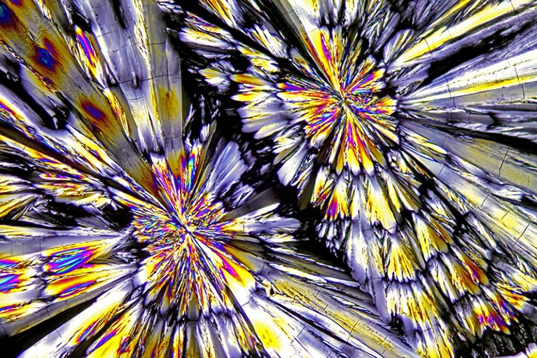
A light micrograph of Ketamine crystals.

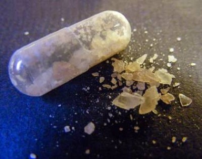
Crystals of ketamine, which is growing in popularity as a treatment for depression and anxiety.Credit: M. I. Walker/Science Photo Library

The drug ketamine is enjoying a second life. First developed as an anaesthetic that was used widely by US battlefield surgeons during the Vietnam war, it is growing in popularity for treating depression and other mental-health conditions. And this week, the drug got its highest-profile endorsement yet.

In an interview with US journalist Don Lemon that was released online Monday, Elon Musk, founder of SpaceX and head of social-media platform X (formerly Twitter), spoke of his own experiences of using the drug to manage what he called a “negative chemical state” similar to depression. Musk said he has a prescription for the drug from “a real doctor” and uses “a small amount once every other week or something like that.” His comments follow the fatal drowning of Friends actor Matthew Perry in October last year, which an investigation blamed on acute effects of the drug.

So what is ketamine, how is it used and is it safe? Nature spoke to three specialists in the field to find out.

How is ketamine used against depression?

It’s complicated. Approved as an anaesthetic by the US Food and Drug Administration in 1970, the drug was delivered intravenously to people undergoing surgery. Ketamine is often still given that way for depression. That demands supervision — typically a person attends a private clinic and is monitored by an anaesthetist as well as the prescribing psychiatrist and support staff.

Because it’s long out of patent, there’s little commercial interest in developing new versions of ketamine. Some companies are trying to package it into more convenient oral lozenges, but that’s a challenging formulation.

“The problem with ketamine is if you take it orally, by and large it doesn’t get through to the system because it’s got low bioavailability,” says Allan Young, a consultant psychiatrist at King’s College London who studies mood disorders.

In 2019, the FDA approved a nasal spray that contains a potent ketamine derivative called esketamine as a treatment for severe depression. But it’s more expensive than standard ketamine, so many doctors simply prescribe the tried and tested anaesthetic off-label. They are doing so more often: data suggests that ketamine prescriptions in the United States rose more than fivefold from 2017 to 2022.

Does it work?

Ketamine faces a high bar, as it tends to be given to people with more severe depression who have already tried standard treatments but received little or no benefit. But even in people who did not respond to other drugs, ketamine can prove highly effective.

“We see about 50 to 60% of people having a very clinically meaningful response,” says Joshua Rosenblat, a staff psychiatrist and clinician-researcher at Toronto Western Hospital, Canada. “There’s been over 40 clinical trials now supporting the use of ketamine for depression. For the most part, they’ve just been very, very consistent that there’s a rapid and robust antidepressant effect.”

“We think of ketamine and electroconvulsive therapy (ECT) as quote-unquote big guns that we save for more severe cases,” he adds. (Because of its potency as an anti-depressant, when ketamine first emerged some psychiatrists referred to the drug as “liquid ECT”).

How does it work?

As is true for many psychiatric medicines, scientists aren’t sure how ketamine works at the molecular level in the brain to lift depression. Its benefits seem to come from triggering what psychiatrists call a dissociative state.

People who take it “might feel removed from their body, feeling like they’re in a dreamlike state or like they’re floating,” Rosenblat says.

His clinic typically offers those with treatment-resistant depression an acute course of four to six ketamine treatments over two to four weeks. For those patients who respond, the programme switches to a maintenance schedule of a treatment every two to four weeks — which fits with how often Musk said he takes the drug.

“It’s a very important alternative strategy because it works so differently to conventional medicines,” says David Nutt, a neuropsychopharmacologist at Imperial College London.

Is it used beyond depression?

Although esketamine is approved for some forms of depression, ketamine isn’t licensed in the United States as a treatment for psychiatric conditions. But its success against depression is encouraging researchers and even doctors to investigate its effects for other patients.

“When you get an effective treatment in psychiatry, you tend to look out from the initial indication, to look for benefit,” Young says. His team is studying whether it can help with anorexia. Others are looking at the effects on people with alcohol addiction, anxiety disorders, obsessive-compulsive disorder, and post-traumatic stress disorder (PTSD).

“I don’t prescribe for those indications because I don’t think the evidence is sufficient,” Rosenblat says. “But certainly there’s a lot of prescribers who do. I think it’s really grown in popularity for PTSD.”

Is it safe?

Regulations and medical best-practice say that ketamine should only be administered under the proper conditions, which include supervision for several hours afterwards. Some people receiving the drug for depression do react badly, even at relatively low doses.

“It’s very rare for people to have full perceptual disturbances in terms of hallucinations,” Rosenblat says. “It’s more distortion of time and space and colours and sounds.”

Some people he has treated have entered a very unpleasant state that ketamine users call a K-hole. “You feel like you’re dying, like everything goes black,” he says. “It can be distressing. And so we have staff that are trained in grounding people that sit next to them.”

Not everyone who takes ketamine receives that help. Some clinics and doctors administer the drug unsupervised. And then there’s illegal recreational use, which many see as a growing problem because the drug severely affects judgement.

“There are quite a few documented deaths from ketamine,” Nutt says. “Never take it alone or where you can be vulnerable. Not near water, as [Matthew] Perry did, or outside on a cold night admiring the stars.”

[ad_2]

Source Article Link

Categories
Life Style

First pig liver transplanted into a person lasts for 10 days

[ad_1]

A shopkeeper holds a Bama miniature pig in a cafe offering customers relaxing access to the pet pigs in a shopping mall in Shanghai, China.

Organs from genetically modified pigs could help keep patients alive while they are awaiting a human donor.Credit: Feature China/Future Publishing via Getty

In a milestone for the transplantation of animal organs into people, a 50-year-old clinically dead man in China has become the first person to receive a liver from a pig. With consent from the man’s family, researchers stitched the organ, from a genetically engineered miniature pig, to the man’s blood vessels, where it remained for ten days. It has been surgically removed today, says Dou Kefeng, one of the surgeons who led the transplant at Xijing Hospital of the Air Force Medical University in Xi’an. “Our study has just been terminated, and the colour and texture of the pig liver [transplant] are generally normal.”

The procedure was intended to test whether genetically modified pig organs could one day be used to supply hospitals for transplants. In China alone, hundreds of thousands of people experience liver failure every year, but only around 6,000 received a liver transplant in 2022. In the past few years, surgeons in the United States have transplanted pig hearts into two living people, and transplanted hearts and kidneys to several people declared dead because they lack brain function.

The Xijing surgeons say the pig liver secreted more than 30 millilitres of bile every day, a sign that it was functioning.

Researchers who specialize in transplanting animal organs into people, known as xenotransplantation, are eager to see more details about the procedure’s safety and functional benefits, and to learn from the work.

“This is a really exciting study,” says Ping Li, a transplant researcher at Indiana University School of Medicine in Indianapolis.

Important insights

The surgery marks the first time a pig liver has been transplanted into a human. However, in January, a team led by transplant surgeon Abraham Shaked at the University of Pennsylvania in Philadelphia connected a clinically dead person to a genetically modified pig’s liver located outside their body. The organ circulated the person’s blood for three days.

It is “heart-warming” to see researchers pursuing xenotransplantation all over the world, says Muhammad Mohiuddin, the surgeon and researcher who led the pig-heart transplants in living people. “It’s an expensive process, but it has a huge amount of potential,” says Mohiuddin, who is at the University of Maryland School of Medicine in Baltimore and is president of the International Xenotransplantation Association.

Luhan Yang, chief executive of Qihan Biotech in Hangzhou, China, which is developing gene-edited pigs as a source for organs, says she expects more xenotransplants in clinically dead people or — for compassionate reasons — in terminally ill people in the United States, China and Europe in the coming years.

The Chinese study will offer important insights into whether pig-liver transplants can keep people alive, even just for a few days, says David Cooper, a xenotransplant immunologist at Massachusetts General Hospital in Boston.

Ten days

On 10 March, the Xijing Hospital team, including Dou, Tao Kaishan and Wang Lin transplanted a pig liver weighing 700 grams into the donor, who lacked cognitive function. The surgery took roughly nine hours to perform. The donor received a daily regimen of immunosuppressive drugs, and his original liver was left in place.

The liver came from a Bama miniature pig (Sus scrofa domestica) bred by the company Clonorgan Biotechnology in Chengdu, China. It contained six genetic modifications, says Wang. These deactivated three genes for proteins found on the surface of pig cells and introduced three genes for human proteins, to prevent the donor from rejecting the pig organ.

Dou says the pig was bred in a specialized pathogen-free facility and tested negative for about a dozen pathogens, including Streptococcus suis, the type-2 strain of Mycoplasma pneumoniae and porcine cytomegalovirus. So far, he has not seen signs of an immediate form of organ rejection and the liver is producing bile. “This is encouraging,” says Cooper.

The researchers have also taken daily blood samples and liver biopsies and will assess immune response, infection risk and liver function in detail. “We’re having a pathologist evaluate if there’s acute rejection,” says Dou.

The surgery was approved by the recipient’s family and several university committees, says Wang. “It has been strictly carried out according to relevant national and international regulations.”

Temporary fix?

The researchers plan to repeat the procedure in another clinically dead person later this year — and next time they will remove the person’s existing liver.

Mohiuddin points out that although clinically dead people are a useful model for assessing the viability of xenotransplantation in living people, that usefulness is limited, because once a person’s brain ceases activity, they undergo hormonal changes. And it isn’t yet clear how long someone with no cognitive function can be maintained on a ventilator and with a donated pig organ, he says. The longest documented case was two months, which involved a pig-kidney transplant.

Shaked also questions whether surgery is necessary for pig livers to be useful to humans. Unlike the heart, which essentially functions as a pump, the liver performs many complex tasks, which makes it particularly difficult to transplant. A pig liver can carry out the liver’s detoxifying and waste-disposal role, but Shaked does not anticipate that it will be able to produce the broad array of proteins required for the human liver’s other functions.

This means that whereas heart and kidney xenotransplants have been touted as possible long-term organ replacements, liver xenotransplants are seen mainly as a short-term fix for people with liver failure. They could enable a person’s existing liver to regenerate, for example after damage caused by alcohol or drug consumption, or could buy time while waiting for a human liver donor.

As a result, Shaked and his team chose to avoid operating: they hooked up an external pig liver to the recipient using blood-carrying tubes. But Dou says his team’s goal is organ replacement. He adds that working in a person allows the researchers to collect a lot more data, including information on immunology and physiological changes.

Yang says she hopes the team will publish detailed insights about the transplantation in peer-reviewed publications, to help determine which approach is more feasible.

In the meantime, Shaked hopes to exchange notes with the Chinese team. “I’d love to hear more about what they did. It’s fantastic.”

[ad_2]

Source Article Link

Categories
Life Style

What Putin’s election win means for Russian science

[ad_1]

Vladimir Putin behind a lectern marked with a golden eagle crest.

Vladimir Putin spoke at an event marking the 300th anniversary of the Russian Academy of Sciences.Credit: Getty Images

Russian President Vladimir Putin has secured a fifth term in office, claiming a landslide victory in the country’s presidential election on 18 March. Election officials say he won a record 87% of votes. This outcome came as a surprise to no one, and many international leaders have condemned the vote as not being free or fair.

Researchers interviewed by Nature say that another six years of Putin’s leadership does not bode well for Russian science, which has been shunned globally in response to the country’s ongoing invasion of Ukraine, and is on precarious ground at home. Those still in Russia must choose their words carefully: as one scientist, who wishes to remain anonymous, put it, “business as usual” now includes possible prison time for offhand comments.

Publicly, Putin’s government is a big supporter of research. In early February, at a celebration of the 300-year anniversary of the Russian Academy of Sciences, Putin bolstered the academy’s role, effectively reversing parts of a sweeping reform that limited its autonomy he oversaw in his third term. And at the end of last month, he signed an update to the 2030 national science and technology strategy, which calls for funding for research and development to double to 2% of gross domestic product, and stresses an increased role for applied science amid “sanctions pressure”.

Despite being made before the election, these big announcements were framed not as campaign promises but as top-down directives, says Irina Dezhina, an economist at the Gaidar Institute for Economic Policy in Moscow. “The fact that it was set in motion back then implies no one really expected any changes at the helm.”

Fractured landscape

Although domestic support for Russian science, which remains mostly state-funded, appears to be strong, many collaborations with countries in the West have broken down since the invasion of Ukraine, prompting a shift to new partners in India and China.

After intense internal discussions, CERN, the European particle-physics powerhouse near Geneva, Switzerland, voted in December 2023 to end ties with Russian research institutions once the current agreement expires in November this year. And the war has severely disrupted science in the Arctic, where Russia controls about half of a region that is particularly vulnerable to climate change. A study1 this year gave a sense of how collaborative projects could be affected by losing Russian data: excluding Russian stations from the International Network for Terrestrial Research and Monitoring in the Arctic causes shifts in project results that are in some cases as large as the total expected impact of warming by 2100.

Reports also suggest that political oppression combined with the threat of military draft have led to a ‘brain drain’ among scientists. Getting an accurate headcount is challenging, but a January estimate by the Latvia-based independent newspaper Novaya Gazeta Europe, based on researchers’ ORCID identifiers, says at least 2,500 researchers have left Russia since February 2022.

Researchers who stayed in Russia have had to contend with serious supply-chain disruptions as well as personal risks. And international sanctions on Russia might have hit even the most productive scientists: according to a January 2024 paper co-authored by Dezhina, which surveyed some of the most published and cited Russian researchers, three out of four of them report at least some fallout from sanctions, mostly economic ones2.

Russia’s isolation has particularly affected the medical sciences, because it means that international clinical trials are no longer held there, says Vasily Vlassov, a health-policy researcher at the Higher School of Economics University in Moscow. He fears that being cut off from the global community will erode Russia’s expertise in this fast-moving and technically complex field: “It’s a problem we have yet to fully appreciate.”

Researchers in the social sciences and humanities are less dependent on overseas partners, but they are affected by increasingly nationalist ideology, says a Russian researcher who asked to remain anonymous. When reviewing articles for publication in Russian journals, the researcher says, they are seeing an increasing number of submissions blaming problems in research and higher education on ‘the collective West’, a common propaganda term. “It’s everywhere, and it’s poisoning minds.”

Uncertain future

The election outcome serves as a reminder of the ongoing war and the openly totalitarian environment in Russia, says Alexander Kabanov, chief executive of the Russian-American Science Association, a US-based non-profit organization. “We are still dealing with an ongoing disaster,” he says.

Yet the impacts of sanctions on Russian science are beginning to fade from public consciousness in other countries. Pierre-Bruno Ruffini, who studies science diplomacy at Le Havre University-Normandy in Le Havre, France, says that academic sanctions and their consequences have “rapidly and completely disappeared” from discussions in the French research community. Dezhina agrees, and adds that, in her experience, even cooperation between individual scientists, once seen as a promising workaround for institutional bans, is on the decline.

Researchers in exile are working on an alternative to the state’s vision of the future for Russia and national science. A policy paper published earlier this month by Reforum, a European project that aims to create a “roadmap of reforms for Russia”, presents a to-do list for revitalizing Russian research. Three out of five of the tasks listed focus on bringing it back into the international fold. Olga Orlova, a science journalist who wrote the policy paper, thinks that scientists in Russia have a part in building that future.

“They shouldn’t be afraid of the change — they should be working for it,” she says.

[ad_2]

Source Article Link

Categories
Life Style

AI & robotics briefing: LLMs harbour hidden racism

[ad_1]

Hello Nature readers, would you like to get this Briefing in your inbox free every week? Sign up here.

A laptop user is typing on the Google Bard AI chatbot webpage.

Some models are more likely to associate African American English with negative traits than Standard American English.Credit: Jaap Arriens/NurPhoto via Getty

Some large language models (LLMs), including those that power chatbots such as ChatGPT, are more likely to suggest the death penalty to a fictional defendant presenting a statement written in African American English (AAE) compared with one written in Standardized American English. AAE is a dialect spoken by millions of people in the United States that is associated with the descendants of enslaved African Americans. “Even though human feedback seems to be able to effectively steer the model away from overt stereotypes, the fact that the base model was trained on Internet data that includes highly racist text means that models will continue to exhibit such patterns,” says computer scientist Nikhil Garg.

Nature | 5 min read

Reference: arXiv preprint (not peer-reviewed)

A drug against idiopathic pulmonary fibrosis, created from scratch by AI systems, has entered clinical trials. Researchers at Insilico Medicine identified a target enzyme using an AI system trained on patients’ biomolecular data and scientific literature text. They then used a different algorithm to suggest a molecule that would block this enzyme. After some tweaks and laboratory tests, researchers had a drug that appeared to reduce inflammation and lung scarring. Medicinal chemist Timothy Cernak says he was initially cautious about the results because there’s a lot of hype about AI-powered drug discovery. “I think Insilico’s been involved in hyping that, but I think they built something really robust here.”

Chemical & Engineering News | 4 min read

Reference: Nature Biotechnology paper

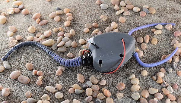
Researchers built a pleurocystitid robot to investigate how the ancient sea creature moved. Pleurocystitids lived 450 million years ago and were probably one of the first echinoderms (animals including starfish and sea urchins) that could move from place to place using a muscular ‘tail’. The robot moved more effectively on a sandy ‘seabed’ surface when it had a longer tail, which matches fossil evidence that pleurocystitids evolved longer tails over time.

Ars Technica | 5 min read

Reference: PNAS paper

Image of Rhombot robot testbed inspired by anatomy of pleurocystitid.

The tail of the pleurocystitid replica (nicknamed ‘Rhombot’) was built out of wires that contract in response to electrical stimulation to simulate the flexibility and rigidity of a natural muscular tail.(Carnegie Mellon University – College of Engineering)

Features & opinion

Scientists hope that getting AI systems to comb through heaps of raw biomolecular data could reveal the answer to one of the biggest biological questions: what does it mean to be alive? AI models could, with enough data and computing power, build mathematical representations of cells that could be used to run virtual experiments — as well as map out what combination of biochemistry is required to sustain life. Researchers could even use it to design entirely new cells, that, for example, can explore a diseased organ and report on its condition. “It’s very ‘Fantastic Voyage’-ish,” admits biophysicist Stephen Quake. “But who knows what the future is going to hold?”

The New York Times | 9 min read

The editors of Nature Reviews Physics and Nature Human Behaviour have teamed up to explore the pros and cons of using AI systems such as ChatGPT in science communication. Apart from making up convincing inaccuracies, write the editors, chatbots have “an obvious, yet underappreciated” downside: they have nothing to say. Ask an AI system to write an essay or an opinion piece and you’ll get “clichéd nothingness”.

In Nature Human Behaviour, six experts discuss how AI systems can help communicators to make jargon understandable or translate science into various languages. At the same time, AI “threatens to erase diverse interpretations of scientific work” by overrepresenting the perspectives of those who have shaped research for centuries, write anthropologist Lisa Messeri and psychologist M. J. Crockett.

In Nature Reviews Physics, seven other experts delve into the key role of science communication in building trust between scientists and the public. “Regular, long-term dialogical interaction, preferably face-to-face, is one of the most effective ways to build a relationship based on trust,” notes science-communication researcher Kanta Dihal. “This is a situation in which technological interventions may do more harm than good.”

Nature Reviews Physics editorial | 4 min read, Nature Human Behaviour feature | 10 min read & Nature Reviews Physics viewpoint | 16 min read

Technology journalist James O’Malley used freedom-of-information requests to unveil how one of London’s Underground stations spent a year as a testing ground for AI-powered surveillance. Initially, the technology was meant to reduce the number of people jumping the ticket barriers, but it was also used to alert staff if someone had fallen over or was spending a long time standing close to the platform edge. Making every station ‘smart’ would undoubtedly make travelling safer and smoother, argues O’Malley. At the same time, there are concerning possibilities for bias and discrimination. “It would be trivial from a software perspective to train the cameras to identify, say, Israeli or Palestinian flags — or any other symbol you don’t like.”

Odds and Ends of History blog | 14 min read

Image of the week

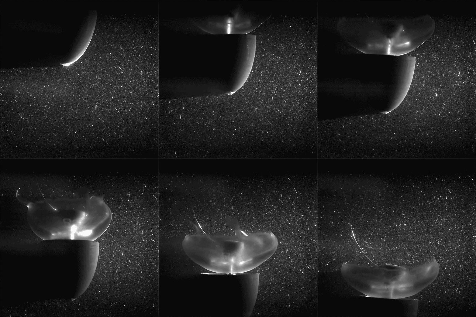
An animated sequence of the Jellyfish biohybrid robot swimming with an attached hemi-ellipsoid forebody.

Simon R Anuszczyk and John O Dabiri/Bioinspir. Biomim. (CC BY 4.0)

A 3D-printed ‘hat’ allows this cyborg jellyfish to swim almost five times faster than its hat-less counterparts. The prosthesis could also house ocean monitoring equipment such as salinity, temperature and oxygen sensors. Scientists use electronic implants to control the animal’s speed and eventually want to make it fully steerable, in order to gather deep ocean data that can otherwise only be obtained at great cost. “Since [jellyfish] don’t have a brain or the ability to sense pain, we’ve been able to collaborate with bioethicists to develop this biohybrid robotic application in a way that’s ethically principled,” says engineer and study co-author John Dabiri. (Popular Science | 3 min read)

Reference: Bioinspiration & Biomimetics paper

Quote of the day

Machine-learning engineer Rick Battle says that chatbots’ finicky and unpredictable performance depending on how they’re prompted makes sense when thinking of them as algorithmic models rather than anthropomorphized entities. (IEEE Spectrum | 12 min read)

[ad_2]

Source Article Link

Categories
Life Style

how a small team created the largest mouse-embryo atlas so far

[ad_1]

Building an atlas of all the cell types that make up the body typically requires multinational collaborations and massive budgets. But a technique that can analyse the genetic activity of hundreds of thousands of individual cells at a time has allowed one small team to produce a time-lapse atlas of an embryonic mouse’s cells over ten days of development. The atlas, which was created by three researchers in one year for approximately US$370,000, could help scientists to understand how stem cells turn into specific cell types, how organs develop and even how the body changes just after it is born.

The study, which was published in Nature in February1, “is impressive at many levels, both the scale of what they achieved and how they achieved it”, says Bertie Göttgens, a stem-cell biologist at the University of Cambridge, UK, who was not involved in the study.

Geneticist Jay Shendure at the University of Washington in Seattle doesn’t normally study mouse development. His laboratory is known for establishing molecular-biology techniques, including one called sci-RNA-seq3 that allows researchers to survey the assemblage of messenger RNA (mRNA), known collectively as the transcriptome, in individual cells.

Instead of looking at whole cells, which would be difficult to keep intact through the process, scientists grind up a sample — in this case, a whole mouse embryo — and isolate its cell nuclei. They split these nuclei into individual dishes and add a different molecular tag to the mRNA in each dish. Next, they combine the nuclei, separate them again, mark each dish with a new tag and repeat. Eventually, each nucleus acquires a unique collection of tags — a molecular barcode — that the researchers can use to determine which tags define the cell’s transcriptome. They can then sequence these cells’ mRNA and construct a ‘tree’ that models how one cell type can turn into another, doing so across multiple animals of different ages on the basis of the genes they express.

Missing moments

Two of Shendure’s lab members, postdoc Chengxiang Qiu and research scientist Beth Martin, decided to demonstrate sci-RNA-seq3 by charting the single-cell transcriptomes of embryonic mice during the animals’ roughly 19-day gestation period. At first, they collected embryos every 24 hours over a 5-day period, but the transcriptomes changed so much between time points that it was difficult to follow how stem cells turned into specific cell types over time2. Shendure likens it to a video that is missing too many frames: more like a stop-motion animation than a smooth progression.

So Martin and Qiu partnered with research scientist Ian Welsh at the Jackson Laboratory, a research institute and mouse-breeding facility in Bar Harbor, Maine. Welsh painstakingly collected 83 mouse embryos at 2–6-hour intervals over 10 days of gestation, from the point at which organs start to develop up until just after the animal’s birth. Welsh snap-froze the embryos and sent them to Seattle, where Martin collected single-cell transcriptomes. Qiu then mapped the data into trees that show when and how each of 190 cell types — liver or bone-marrow cells, for instance — originates in an embryo.

To flesh out the tree, the researchers integrated their data, which began eight days into gestation, with existing work from Shendure’s team and others that had mapped the transcriptomes of these and younger embryos. This added another 110,000 cells to the mix, and these data formed the tree’s ‘roots’, allowing the researchers to follow the branching of early stem cells into specific types seen in the older embryos.

The resulting atlas, containing the transcriptomes of mice across 45 time points, is now available for developmental biologists to study in more depth. With 12.4 million cells, it is the largest mouse-embryo atlas so far and is nearly one-quarter the size of the cell data collected by the Human Cell Atlas collaboration, which comprises 700 labs attempting to map all of the cells in the human body.

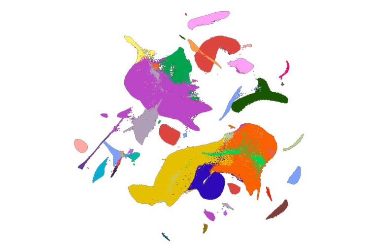
UMAP representation of an atlas of mouse cells during prenatal development

A 2D visualization of the mouse-atlas data set, with colours corresponding to 26 major cell types.Credit: C. Qiu et al./Nature

“It’s a fantastic resource for the community,” says cellular geneticist and Human Cell Atlas co-founder Sarah Teichmann at the Wellcome Sanger Institute in Hinxton, UK. Teichmann points out that there is still work to be done on the mouse atlas. Some time points have more complete transcriptomes than others, and the researchers have not yet separated mice by sex to look at those differences. But she says it will enable a number of studies, including the ability to compare mouse and human development. Shendure says he and his team plan to create single-cell atlases of juvenile and adult mice from conception to death.

Stress effects

Although Shendure and his group aim to let others conduct in-depth biological analyses of the data, they did note two phenomena in their paper. The point at which the transcriptome changed most dramatically, they found, was in the hour just after birth, which Shendure calls “the most stressful moment in your life”. Some of those differences were expected — lung and fat cells changed activity to cope with being outside the uterus, for instance — but other changes are still unclear.

Pure luck led them to another finding. To get the timing just right, Welsh typically delivered the mice by caesarean section. But one day, he returned from lunch to an unexpected nest of newborn pups. Martin processed the mice anyway and found that their transcriptomes were significantly different from those of mice born by caesarean section. Those differences could explain the variation in health outcomes seen between people who were born by these two methods, the researchers say.

Yonatan Stelzer, an epigeneticist at the Weizmann Institute of Science in Rehovot, Israel, says the study is encouraging for future efforts to map the cells of individual organs or tissues. The next step for embryos, he says, will involve not only studying how cells develop over time, but also following them through space in 3D, tracking how they split and move to form a whole mouse. Future research, he adds, could also investigate questions such as how two cells with similar transcriptomes end up with different fates to become the right or left eye, for instance. “We’re still far from solving the entire embryonic puzzle,” he says.

[ad_2]

Source Article Link

Categories
Life Style

Rotating curved spacetime signatures from a giant quantum vortex

[ad_1]

To experimentally realize a curved spacetime such as a black hole requires a specific relative motion between the excitations and the background medium. One-dimensional supersonic flow, the archetypal example of an acoustic black hole, provides a platform for observations of Hawking radiation in both classical20,21 and quantum fluids9,10,22. More complex phenomena such as Penrose superradiance require rotating geometries realizable in two spatial dimensions, for example, by means of a stationary draining vortex flow12,23. Classical fluid flow experiments have demonstrated the power of the gravity simulator programme, realizing superradiant amplification of both coherent11,24 and evanescent waves25, as well as quasinormal mode oscillations26, a process intimately connected to black hole ringdown27.

Here we investigate related phenomena in the limit of negligible viscosity in superfluid 4He (called He II). Its energy dissipation is dependent on temperature and can be finely adjusted across a wide range. At 1.95 K, at which our experiments take place, its kinematic viscosity is reduced by a factor of 100 compared with water28 and the damping is dominated by thermal excitations collectively described by the viscous normal component28,29 that constitutes approximately half of the total density of the liquid. Moreover, He II supports the existence of line-like topological defects called quantum vortices. Each vortex carries a single circulation quantum κ ≈ 10−7 m2 s−1 and forms an irrotational (zero-curl) flow field in its vicinity29. Owing to this discretization, a draining vortex of He II can manifest itself only as a multiply quantized (also known as giant) vortex or as a cluster of single quantum vortices. Such vortex bundles exhibit their own collective dynamics and can even introduce solid-body rotation30 at length scales larger than the inter-vortex distance, adding complexity to the study of quantum fluid behaviour. As the realization of curved spacetime scenarios requires an irrotational velocity field1,31, it is critical to confine any rotational elements into a central area, that is, the vortex core. However, alike-oriented vortices have a tendency to move apart from each other, which poses a limitation on the extent of the core one can stabilize in an experiment. On the other hand, recent findings show that mutual friction29 between quantum vortices and the normal component contributes to the stabilization of dense vortex clusters32.

The vortex induces a specific velocity field within the superfluid, which affects the propagation of small waves on its surface. In particular, low-frequency excitations perceive an effective acoustic metric3,4

$${g}_{ij}\propto \left(\begin{array}{rc}-{c}^{2}+{v}^{2} & -{\bf{v}}\\ -{\bf{v}} & {{\mathbb{1}}}_{2\times 2}\end{array}\right),$$

(1)

in which c denotes their propagation speed and \({\bf{v}}(r,\theta )={v}_{r}\widehat{{\bf{r}}}+{v}_{\theta }\widehat{{\boldsymbol{\theta }}}\) indicates the velocity field at the interface (we assume that the superfluid and normal velocity fields are equal, in line with other mechanically driven flows of He II (refs. 33,34)). Although this description fails in the high-frequency regime owing to dispersion, it is well known that the curved spacetime phenomenology persists for these excitations24,26,35. Altogether, the above properties suggest that an extensive draining vortex of He II is a feasible candidate for simulations of a quantum field theory in curved spacetime.

We realized this flow in cylindrical geometry that is built on the concept of a stationary suction vortex36 (see Methods for a detailed description). The central component of our set-up is a spinning propeller, which is responsible for establishing a continuous circulating loop of He II, feeding a draining vortex that forms in the optically accessible experimental zone. At small propeller speeds, we observe a depression on the superfluid interface (Fig. 1a), but as the speed increases, this depression deepens and eventually transforms into a hollow vortex core extending from the free surface to the bottom drain (Fig. 1b). The parabolic shape of the free surface in the former regime is consistent with solid-body rotation, which corresponds to a compact, polarized cluster of singly quantized vortices (called solid core) that forms under the finite depression. The hollow core can instead absorb individual circulation quanta and behave like a multiply quantized object37. To minimize the rotational flow injected by the spinning propeller into the experimental zone, we devised a unique recirculation strategy based on a purpose-built flow conditioner (see Methods) that promotes formation of a centrally confined vortex cluster instead of a sparse vortex lattice. However, the exact dynamics of individual quantum vortices, as well as their spatial distribution in the experiment, calls for future investigations. State-of-the-art numerical models38 account for the motion of vortex lines coupled to the superfluid and normal velocity fields, but fail to dynamically model the interface, which is a pivotal element in our system. Previous experimental efforts39,40,41 confirmed that a draining vortex in He II carries macroscopic circulation but lacked spatial resolution required to investigate central confinement of rotational components. In this regard, cryogenic flow visualization42 provides sufficient resolution. However, this method requires introducing small solid particles into the superfluid, which accumulate along the vortex lines and considerably affect their dynamics43.

Fig. 1: Side views of two distinct configurations of the giant quantum vortex.
figure 1

a, At low propeller frequencies (here 1 Hz), the interface exhibits a discernible depression, and the vortex core beneath takes the form of a compact, polarized cluster of singly quantized vortices (called solid core). b, With the escalation of frequency (here to 2 Hz), a fully formed hollow core emerges, behaving like a multiply quantized object. Dark vertical stripes in the background provide contrast to the imaged interface. A simplified sketch of this interface (white lines) helps to identify these regimes in later figures. Scale bar, 10 mm.

The above limitations compelled us to propose an alternative, minimally invasive method to examine the vortex flow and extract macroscopic flow parameters that exploit the relative motion occurring between interface waves and the underlying velocity field. The corresponding dispersion relation for angular frequencies ω and wave vectors k reads35

$${(\omega -{\bf{v}}\cdot {\bf{k}})}^{2}=F(\parallel {\bf{k}}\parallel ),$$

(2)

in which F denotes the dispersion function. By solving equation (2), we find (see Methods) that the spectrum of interface modes gets frequency shifted and the velocity field can be inferred from these shifts44. Therefore, we redirect our attention towards precise detection of small waves propagating on the superfluid interface.

We identified that the adapted Fourier transform profilometry17,18 is well suited to our needs, as it is capable of resolving a fluid interface with sufficient and simultaneous resolution in both space and time. This powerful technique consists of imaging the disturbed interface against a periodic backdrop pattern. This way, we resolve height fluctuations of said interface (Fig. 2a) with sensitivity up to approximately one micrometre. Owing to symmetries of the flow, the waves exhibit two conserved quantities: frequency f and azimuthal number m. The latter parameter counts the number of wave crests around a circular path, with positive or negative values of m corresponding to wave patterns co-rotating or counter-rotating with the central vortex.

Fig. 2: Superfluid interface reconstruction and wave analysis.
figure 2

a, Snapshot of the free helium surface depicts height fluctuations representing micrometre waves excited on the superfluid interface. Grey areas mark the positions of the central drain (radius 5 mm) and the outer glass wall (radius 37.3 mm). be, Examples of different azimuthal modes |m| (m counting the number of wave crests or troughs around a circular path) extracted from panel a by a discrete Fourier transform. Wave amplitudes are rescaled for better visibility. f,g, Two-dimensional wave spectra obtained by transforming angle and time coordinates, for radii of 11.2 mm (panel f) and 22.1 mm (panel g). These radii are marked in panel a by coloured circles. Absence of excitations in low-frequency bands (below the coloured lines) can be understood through the solution of equation (2). The corresponding theoretical predictions of the minimum frequency permissible for propagation for the given radii can be matched with experimental observations (yellow and red lines).

These spatial patterns (or modes) can be retrieved from the height-fluctuation field by a discrete Fourier transform. For example, by transforming with respect to the angle θ, we can single out individual azimuthal modes (Fig. 2b–e). To study wave dynamics in time, we must also transform the temporal coordinate and inspect the resulting two-dimensional spectra, showcased in Fig. 2f,g for two distinct radii. Notable high-amplitude signals in the m = ±1 bands are exclusively a consequence of how mechanical vibrations of the set-up imprint themselves on our detection method. Of physical interest are modes with higher azimuthal numbers. These excitations, observed in both solid-core and hollow-core regimes, represent micrometre waves excited on the interface. In the steady state, the waves dissipate their energy, in part by viscous damping and in part by scattering into the draining core of the vortex45. Although this is balanced by the stochastic drive originating from the fluid flow and/or aforementioned mechanical vibrations, we notice that only a certain region of the spectral space (m, f) is populated with excitations, a feature that varies when examining smaller (Fig. 2f) and larger (Fig. 2g) radii. We observe that only some high-frequency (equivalent to high-energy) waves have the capability to propagate on the interface. Through the solution of equation (2), we can pinpoint the minimum frequency, fmin, permissible for propagation for the given radius, azimuthal number and background velocity (see Methods) and, in line with the methodology introduced above, we exploit this particular frequency to extract the underlying velocity field, as we now describe. We search the parameter space produced by two velocity components (vr, vθ) and determine values that produce the best match between fmin and the lowest excited frequency in the experimental data across several azimuthal modes (coloured lines in Fig. 2f,g). By carrying out this procedure for every examined radius, we can reconstruct the velocity distribution in the draining vortex flow.

We conducted these reconstructions across several vortex configurations distinguished by the drive (propeller) frequency. For all instances, vr approximates zero within the limits of our resolution. Although seemingly paradoxical, this outcome results from a complex boundary-layer interaction and is in agreement with earlier findings in classical fluids46. Therefore, interface waves engage with an almost entirely circulating flow characterized by a specific radial dependence of vθ (coloured points in Fig. 3a). Overall, the results are consistent with

$${v}_{\theta }(r)=\varOmega r+\frac{C}{r},$$

(3)

indicated in Fig. 3a by coloured lines. The first term represents solid-body rotation with angular frequency Ω, which leaks into the experimental area through the flow conditioner as described above. The second term corresponds to an irrotational flow around a central vortex with circulation C. The related number of circulation quanta confined in its core, NC = 2πC/κ, is shown in Fig. 3b as a function of the drive frequency. Across all instances, the core consists of the order of 104 quanta, a record-breaking value in the realm of quantum fluids. In the solid-core regime, NC can be identified with the number of individual quantum vortices concentrated in the core. However, in the context of a hollow core, NC represents its topological charge. Achieving circulation values separated from the elementary quantum κ by four orders of magnitude allows the quantization of circulation to be disregarded, leaving the vortex effectively classical. This unprecedented realization of a giant quantum vortex flow represents a distinctive instance of a quantum-to-classical flow transition in He II (ref. 47).

Fig. 3: Reconstructed velocity distribution and flow parameters.
figure 3

a, Coloured points denote the radial dependence of the azimuthal velocity vθ for six vortex configurations distinguished by the drive (propeller) frequency. Each point is obtained by averaging over a 2.5-mm radial interval. Radial velocity component is approximately zero across all instances. Best fits of vθ(r) (coloured lines) yield the circulation C of the central vortex and the angular frequency Ω of the extra solid-body rotation. b, Number of circulation quanta confined in the vortex core, NC = 2πC/κ, corresponds to the most extensive vortex structures ever observed in quantum fluids. c, The ratio η between Ω and the angular frequency of the drive is less than 2.5% in all cases, suggesting that the velocity field in our system is dominated by the irrotational vortex flow. Vertical error bars in panels a and c denote one standard deviation intervals. Standard deviation intervals of data points in panel b, comparable with the symbol size, are not shown.

The importance of the aforementioned outcomes can be underlined by noting that n-quantized vortices are dynamically unstable13,14. They spontaneously decay into a cluster of n vortices48 as a result of the excitation of a negative energy mode in the multiply quantized vortex core11,48. Nevertheless, dynamical stabilization of giant vortices can be achieved by suitably manipulating the superfluid. Namely, introducing a draining flow and reducing the fluid density at the centre has proven effective in polariton condensates, for vortices with n 100 (refs. 15,16). These results agree with our experiment, in which the reduced density translates into the existence of a hollow core and the draining flow resides in the bulk of the draining vortex.

It is worth noting that larger circulation values around a draining vortex in He II are documented in the literature41. However, therein, the contributions of the vortex core and the solid-body rotation are not distinguished. The second effect may dominate in the reported circulations, as the number of quantum vortices responsible for rotation30 scales with the corresponding angular frequency Ω. Rotation in our experimental zone is notably suppressed. The sparse presence of quantum vortices partially justifies our assumption that normal and superfluid components behave as a single fluid. More importantly, the ratio η between Ω and the angular frequency of the drive does not exceed 2.5% (Fig. 3c), and the velocity field in our system is dominated by the irrotational vortex flow. The core of this vortex must be smaller than 7.6 mm, the smallest investigated radius, because the velocity profiles (Fig. 3a) show no indication of a turning point at small radii.

We can, nonetheless, venture beyond the experimental range by exploring wave dynamics in the radial direction. We restrict our discussion to a particular mode |m| = 8 (Fig. 2d) as a representative of the outlined behaviour. We start by analysing co-rotating (m = 8) modes, shown in Fig. 4a,b for the solid-core and hollow-core structures. In both cases, fmin (red line) denotes an effective potential barrier, preventing waves from reaching the vortex core. Existence of this barrier, together with an outer, solid boundary at 37.3 mm, gives rise to bound states (standing waves), appearing as distinct, striped patterns extending up to 40 Hz. These patterns represent the first direct measurement of resonant surface modes around a macroscopic vortex flow in He II.

Fig. 4: Bound states in co-rotating waves.
figure 4

Fourier amplitudes of interface waves corresponding to m = 8 mode show a characteristic pattern in the radial direction that can be identified with bound states, that is, standing waves between the outer boundary (glass wall) at 37.3 mm and the effective potential barrier (red lines). A simplified but accurate model of the potential (yellow lines) is extended beyond the experimentally accessible range (dashed black lines). a, Solid-core regime. Rescaled amplitudes of four bound states labelled I–IV (blue lines) are shown as a function of radius. Crossing points with the potential barrier are marked by yellow points. b, Hollow-core regime. c, Comparison of bound-state frequencies retrieved from panel a (red points) and their theoretical predictions (black circles). Frequencies of states I–IV are highlighted by blue arrows.

To perform an in-depth examination of selected states (denoted as I–IV), we plot the absolute value of their amplitudes in Fig. 4a. The frequency of state I meets fmin in a crossing point (yellow point) located within the field of view. At large radii, this wave harmonically propagates. However, as it penetrates the barrier, its amplitude exponentially decays in exact analogy with a simple quantum-mechanical model of a particle trapped in a potential well. For higher frequencies, the crossing point moves towards smaller radii (state II), eventually reaching the limit of our detection range. For the highest-frequency states (III and IV), the crossing point is well outside the detection range and we only observe the harmonic part of the signal. Nonetheless, the mere existence and predictability of these states lets us extend the effective potential barrier beyond the observable range.

Specifically, we consider a model of a purely circulating vortex, whose velocity field reads (vr, vθ) = (0, C/r), and extend the experimentally determined potential barrier (red lines in Fig. 4a,b) towards smaller radii (yellow lines). In practice, this model must break down near the vortex core, at which point the spatial distribution of individual quantum vortices becomes relevant. Nonetheless, the frequencies of individual bound states are in excellent agreement with theoretical predictions (see Methods) based on the extended potential barrier (Fig. 4c). This outcome validates the simplified model and allows us to constrain the radius of the core region to approximately 4 and 6 mm, respectively for the solid-core and hollow-core regimes. Confinement of the rotating core beyond the experimental range gains importance when considering the draining vortex flow as a gravity simulator, for example, when searching for initial indications of black hole ringdown.

For this purpose, we focus on counter-rotating (m = −8) modes, depicted in Fig. 5a,b with the effective potential barriers (red lines) and their extensions (yellow lines). The shape of the barrier in the solid-core regime (Fig. 5a) allows the existence of bound states up to approximately 30 Hz. However, this is not the case in the hollow-core regime (Fig. 5b), despite the corresponding circulations only differing within one order of magnitude. Bound states are not formed at all because the effective potential shows a shallow maximum before decreasing towards zero. Dominant excitations in this spectrum, highlighted in Fig. 5c, are modes lingering near this maximum. These excitations, previously identified as ringdown modes of an analogue black hole26, represent the very first hints of this process taking place in a quantum fluid. The radius at which the effective potential crosses the zero-frequency level is related to the analogue ergoregion35, a key feature in the occurrence of black hole superradiance. To directly observe this region in our set-up, further increasing the azimuthal velocity and/or examining the system closer to the vortex core is required.

Fig. 5: Bound states and ringdown modes in counter-rotating waves.
figure 5

Fourier amplitudes of interface waves (same colour scale as in Fig. 4) corresponding to m = −8 mode interact with the effective potential barrier (red lines). Its simplified model (yellow lines) is extended beyond the accessible range (dashed black lines). a, In the solid-core regime, the potential allows existence of bound states, visible up to approximately 30 Hz. b, In the hollow-core regime, no bound states can be retrieved. Instead, we observe dominant excitations lingering near the shallow maximum of the potential (approximately at 8.25 Hz), suggesting the excitation of black hole ringdown modes. c, Inset highlights ringdown mode candidates from panel b, with the effective potential barrier shown as a faint red line.

Our research positions quantum liquids, particularly He II, as promising contenders for finite-temperature, non-equilibrium quantum field theory simulations, marking a transformative shift from already established simulators in curved spacetimes7,8,9,10. The liquid nature of He II arises from an effective, strongly interacting field that complements its weakly interacting counterpart found in, for example, cold atomic clouds. A distinctive advantage presented by He II lies in its flexibility, allowing it to be operated at a fixed temperature, starting just below the superfluid transition, at which He II shows pronounced dissipation. This regime in particular holds immense potential, such as for the mapping to generic holographic theories49. At temperatures below 1 K, the normal component is expected to be an aggregate of individual thermal excitations. This tunability provides the opportunity to investigate a broad range of finite-temperature quantum field theories.

Owing to the capacity of He II to accommodate macroscopic systems, we achieved the creation of extensive vortex flows in a quantum fluid. Notably, the size of the hollow vortex core scales with its winding number and, consequently, system-size constraints may restrict the maximum circulation achievable when implemented in cold-atom or polariton systems alike. Key processes in rotating curved spacetimes, such as superradiance and black hole ringing, can be explored in our current system with minor adjustments to the propeller speed, container geometry or by dynamically varying flow parameters. Our set-up also provides a clear opportunity to investigate rotating curved spacetimes with tunable and genuinely quantized angular momentum, setting it apart from classical liquids. Furthermore, applying these techniques to explicitly time-dependent scenarios allows for the exploration of fundamental non-equilibrium field theory processes. This may involve controlled modulations of first or second sound in the bulk of the quantum liquid, providing a platform for conducting wave-turbulence simulations across various length and temperature scales. This represents a noteworthy advancement beyond the current scope of cold-atom studies50.

[ad_2]

Source Article Link

Categories
Life Style

The warfighter

[ad_1]

Cadence noticed the warfighter on her way to school. He was still there later, sitting in the tiny fenced park, as she walked back home along Sixth Avenue. There was a pigeon watching him, which the police had thought at one time was a good way to disguise a camera. She stopped and sat on the other bench.

“There aren’t any birds,” she said. “You know that, right?”

He was an 800-year-old 30-year-old man and he sat huddled in a large coat with the cheap sheen of post-consumer polyethylene, under which he wore a side-ventilated floral tactical shirt and adaptive camo cargo pants with sagging, empty pockets. She supposed the creases were from being repeatedly flash frozen.

“Why’s the sky grey but it never rains?” he asked.

“Salts.”

“Don’t bullshit me, kid.”

“To keep the sunlight out.”

“That’s fantastic. Thanks for taking care of the place while I was gone.”

“Where’d you fight?”

“Proxima.”

“Where else?”

“Eridani. Lacaille.”

“How old are you?”

“Let’s not do the math.”

“Why’d you come back?”

“The sunsets.”

“Seriously.”

“Come on, give me a break, kid,” he said, getting unsteadily to his feet. “I’ve had a long century.”

She followed him to a coffee kiosk across the street and read the peeling posters on a movie theatre that had closed during a run of Mild Red Pier before she was even born. He paid the machine with a plastic pendant he wore on a bright green spiral wristband, which she was sure also stopped being a thing before she was born. He cracked the lid and sniffed the coffee, then made a face, but he didn’t look back, and she watched as he wandered down the sidewalk and turned a corner, shuffling towards the river.

*****

“He’s from the Cincinnatus,” her father told her. “They returned a month ago. The numina are very interested in him. Few come back these days, and those that do don’t last long.”

“Why?”

“They kill themselves, sweet.”

She looked him up with her headset and found his service record in the National Archives. He was born in analogue New York and enlisted at a time of 77% unemployment, one of thousands to leave behind family and friends to climb into a hydrogel-filled cold tube and accelerate out into the darkness at a fraction of the speed of light. The relationship between temperature and viscosity, she was told, was a mechanism for inertial suspension in Einsteinian space travel, by which time her mind had already started to wander. She heard something about the survival rates back then being only marginally higher than the odds of finding work on the streets of any American city.

*****

She looked for the warfighter in the park the following day, but found only the pigeon perched on a streetlight, head cocked as it scanned passing faces. She threw her apple at the filthy bot, missed, and heard it smack the asphalt of the basketball court.

He was over on the pier, pitching pea gravel out into the clear, shallow Hudson.

“What does a Proximan look like?”

“They’re not called that.”

“What are they called?”

“It translates as something like step-accretion. We called them steppers.”

“What do they look like?”

“Something that comes out the ass end of another stepper.”

“Why did we fight them?”

“Incompatible world views.”

“That’s not a reason.”

“Out there, it’s not about who gets more land, or access to water, or political representation. It’s about the survival of thinking people, period. We’re the only living things out there with a concept of self. Or we were. It’s just one hive mind after another. What does that tell you about the Universe?”

“What?”

“What.”

“What does that tell you about the Universe?” she repeated.

“It tells me I’m sitting out here on the short branch of a very large tree, kid.”

“I have a concept of self.”

“Oh yeah? Did they copy that over from last year’s model?”

“I don’t get a new body every year. I had one as a baby, then this one, and in a few years I’ll be a full adult. Adults get upgraded only every 25 years. It used to be longer, but they make these ones in the maquiladoras.”

“What do you think happens when you’re upgraded?”

“They move my brain from one body to another.”

“Not your brain. Your mind. And they don’t move it, they copy it. And then they put a bullet in the old one.”

“They don’t use bullets.”

“You don’t have a concept of self if everything you think and feel is a mirror image of something else. You have a copy of a concept of self.”

“What’s a mirror?”

“Jesus. Reflective glass. A copy. A duplicate.”

“I’m not a duplicate. There’s only one.”

“Not hanging onto the original doesn’t make a copy any less a copy. People don’t go to the MOMA to see a photograph of Starry Night.”

“What’s a MOMA?” she asked impatiently.

“Go up Sixth to 53rd and hang a right. It’s a peri-peri takeout now.”

*****

Cadence stayed at home the rest of the week studying for her middle-SAT, and when she returned to the pier the warfighter was gone. She watched the narrowband video that showed first landfall on Proxima Centauri b, but the faces were all unfamiliar and the video quality was poor, so she filed it away for later with her unwatched TV shows and the footage from her childhood pets.

She spent the summer on Long Island, and when she returned to school in the fourth quarter she crossed through Washington Square and ambled back down Sixth Avenue. The warfighter was there again in the park. Instead of a pigeon, the numina had deployed a pair of scruffy alleycats, which would provide better stereo.

She sat down on the other bench, but he didn’t recognize her, and after a moment she looked away.

“Why’s the sky grey but it never rains?” asked the two-month-old 800-year-old man.

“Probably smog,” she whispered, and went home.

The story behind the story

Timothy Quinn reveals the inspiration behind The warfighter.

There’s a solution to the Fermi paradox whereby life is commonplace in the Universe, but the complexity and perishability of intelligent, conscious, technological life makes us unique. Intelligence might represent a state of disequilibrium that the Universe does not long abide.

If humanity struggles and wanes, as it does in The warfighter, it might trade evolution for algorithms, war for autocracy, individuality for replicability. To the returning veterans of tomorrow’s abandoned conflicts, Homo sapiens will seem a very different species from that which was left behind hundreds of years before, and it might be difficult to remember exactly what it was that was worth fighting for.

Cadence’s favourite park is the Golden Swan Garden on the Lower East Side of Manhattan, and if you peer over the fence you might find there’s a final matinee screening of Mildred Pierce at the IFC Center.

[ad_2]

Source Article Link

Categories
Life Style

Planet-eating stars hint at hidden chaos in the Milky Way

[ad_1]

Artist's concept of the exoplanet WASP-12b being consumed by its host star.

A star in the process of consuming a planet (artist’s conception).Credit: NG Images/Alamy

Stellar detectives have identified seven stars that recently dined on a rocky planet. The study doubles the number of binary stars known to have consumed a planet, and questions the perception that mature solar systems harbouring Earth-like planets are usually stable.

The findings, published in Nature on 20 March1, show “strong evidence of planet ingestion”, says Jianrong Shi, an astronomer at the National Astronomical Observatories in Beijing. The planets seem to have been eaten during their stars’ relatively stable main-sequence period, adds study co-author Fan Liu, an astronomer at Monash University in Melbourne, Australia.

If this is true, it means these systems have continued to be chaotic long after their formation, with planets disintegrating or falling into their star, says Johanna Teske, an astronomer at the Carnegie Institution for Science in Washington DC. “It’s an inference at this point. We need to look at these systems in more detail,” she says.

Swallowed by stars

Last year, for the first time, astronomers observed a star in the process of eating a planet. But unravelling whether a star has done so in the past is challenging, because planets are tiny compared with their hosts, and their contents soon get diluted.

Different elements absorb and emit light of different wavelengths, so the composition of a star’s surface leaves a fingerprint on the light reaching Earth. But detecting whether a star has eaten a planet is similar to spotting a chocolate chip that’s been swirled into a bowl of vanilla ice cream, says Teske. Stars also vary a lot in their make-up, making it tough to prove that a star has a particular composition because it ingested a planet.

To hunt for planet-eating stars, Liu and his colleagues performed a cosmic-twin study. Using the Gaia space telescope, they found 91 pairs of Sun-like stars nearby in the Milky Way, whose motions suggested that they were both born in the same gas cloud. The stars in such paired systems should have near-identical compositions and their similar lives should rule out many potential causes for discrepancies.

The team then used three ground-based telescopes to study the abundance of 21 elements in the pairs. If there were notable differences between a pair of stars, the researchers looked at whether this could be explained through noise in the data or other sources of variation. For seven pairs, “the difference has to be explained by one [star] ingesting a planet and the other not”, says Meridith Joyce, an astrophysicist at the Konkoly Observatory in Budapest, and a co-author on the paper.

Secret planet-eaters

The study suggests that around 8% of Sun-like star pairs in our region of the Milky Way harbour a planet-eater, says Liu. He adds that this estimate is conservative, because the team considered only stars ingesting rocky planets, whereas other stars might have eaten gaseous Jupiter- or Neptune-like bodies. The method would also have missed cases in which both stars had eaten a planet of similar composition.

Finding clear signs of planet ingestion in billion-year-old stars is “something unexpected”, he says. Astronomers often consider planet-eating to be a feature of a star’s early life, when planetary orbits are unstable and collisions are probable. But these meals must have been relatively recent, in the last few hundred millions of years, or theory suggests the evidence would’ve been undetectable, says Liu. The planets could have met their fate when their eroding atmospheres caused them to spiral inward, or some stars might have captured untethered rogue planets as they flew by, he adds.

Shi says that astronomers should examine these systems to see if any sibling exoplanets remain. The findings should make Earth-dwellers grateful, he says. The diversity of exoplanets has continued to shock astronomers; now it seems that “our Solar System is not only unique, but also undoubtedly peaceful”.

[ad_2]

Source Article Link