Categories
Life Style

Do cutting-edge CAR-T-cell therapies cause cancer? What the data say

[ad_1]

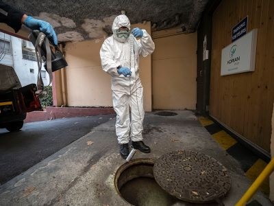
US drug regulators dropped a bombshell in November 2023 when they announced an investigation into one of the most celebrated cancer treatments to emerge in decades. The US Food and Drug Administration (FDA) said it was looking at whether a strategy that involves engineering a person’s immune cells to kill cancer was leading to new malignancies in people who had been treated with it.

Bruce Levine, an immunologist at the University of Pennsylvania Perelman School of Medicine in Philadelphia who helped to pioneer the approach known as chimeric antigen receptor (CAR) T-cell therapy, says he didn’t hear the news until a reporter asked him for comments on the FDA’s announcement.

“Better get smart about it quick,” he remembers thinking.

Although the information provided by the FDA was thin at the time, the agency told reporters that it had observed 20 cases in which immune-cell cancers known as lymphomas had developed in people treated with CAR T cells. Levine, who is a co-inventor of Kymriah, the first CAR-T-cell therapy to be approved, started jotting down questions. Who were these patients? How many were there? And what other drugs had they received before having CAR-T-cell therapy?

The FDA has since documented more cases. As of 25 March, the agency had received 33 reports of such lymphomas among some 30,000 people who had been treated. It now requires all CAR-T therapies to carry a boxed warning on the drug’s packaging, which states that such cancers have occurred. And the European Medicines Agency has launched its own investigation. But many of the questions that Levine had in November remain unanswered. It is unclear how many, if any, of the observed cancers came directly from the manipulations made to the CAR T cells. A lot of cancer therapies carry a risk of causing secondary malignancies, and the treated individuals had received other therapies. As Crystal Mackall, a paediatric oncologist who heads the cancer immunotherapy programme at Stanford University in California, puts it: “Do you have a smoking gun?”

Scientists are now racing to determine whether the cellular therapy is driving these cancers or contributing in some way to their development. From the data available so far, the secondary cancers seem to be a rare phenomenon, and the benefits of CAR T cells still outweigh the risks for most prospective recipients. But it’s an important puzzle to solve so that researchers can improve and expand the use of these engineered cells in medicine. CAR-T-cell treatments were once reserved for people who had few other options for therapy. But the FDA has approved several of these treatments as a relatively early, second-line option for lymphoma and multiple myeloma. And some companies are working to expand the therapy’s repertoire to solid tumours, autoimmune diseases, ageing, HIV and more.

Aric Hall, a haematologist at the University of Wisconsin–Madison, says that despite the enthusiasm for CAR-T therapy, the technology is still new. “I used to joke that for the first ten years there were more review articles about CAR T than there were patients who had been treated by CAR T products,” he says. He adds that the risks might be rare, but as CAR-T therapy moves into a bigger pool of patients who aren’t desperately ill, the calculus could change. “The problem is rare risks become a bigger deal when patients have better options.”

Vector safety

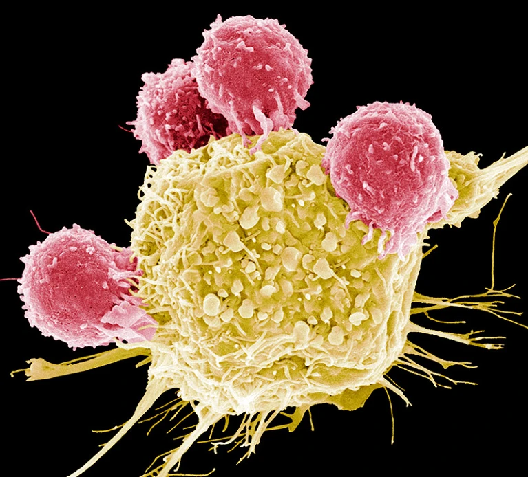
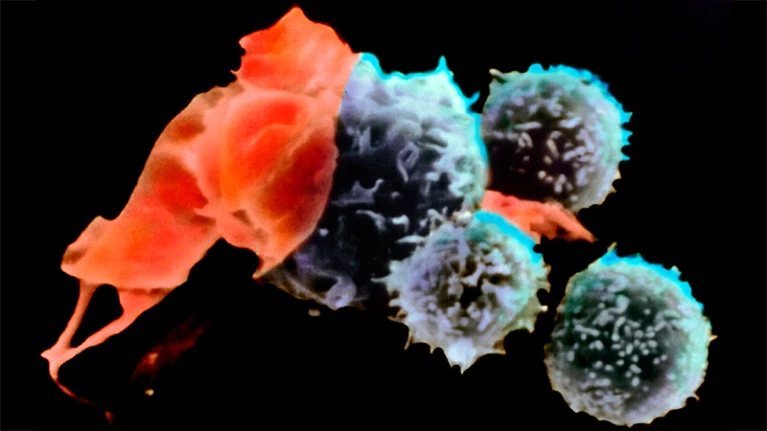
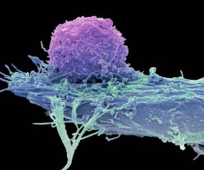
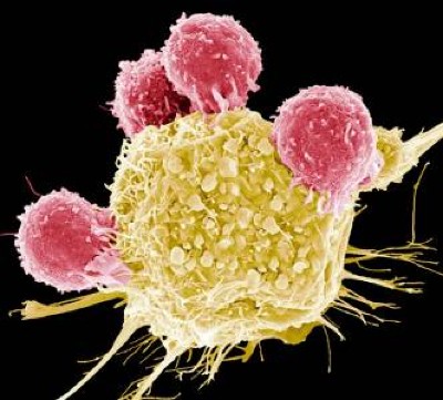
Throughout the development of these blockbuster therapies, researchers had reason to think that CAR T cells could become cancerous. CAR-T therapies are personalized — created from a person’s immune cells. Their T cells are extracted and then genetically modified in the laboratory to express a chimeric antigen receptor — or CAR — a protein that targets the T cell to specific cells that they will kill. T cells sporting these receptors are made to multiply and grow in the lab, and physicians then infuse them back into the individual, where they start battling cancer cells. The six CAR-T-cell therapies currently approved in the United States and Europe target antigens on the immune system’s B cells, so they work only against B-cell malignancies — leukaemias, lymphomas and multiple myeloma. But researchers are aiming to develop CAR-T therapies that work on other kinds of cancer, and for other conditions.

The genetic engineering is the step that creates a risk of malignancy. All six FDA-approved CAR-T therapies rely on a retrovirus — typically a lentivirus, such as HIV, or a gammaretrovirus — to ferry the genetic information into the cell. Scientists remove the parts of the viral genome that allow the virus to replicate, making room for the gene they want the virus vector to carry. Once inside a cell, the virus inserts the gene for the CAR into the cell’s genome. But there isn’t a good way to control exactly where the gene goes. If it slips in near a gene that can promote cancer development and activates it, or if it deactivates a tumour-suppressing gene, that boosts the risk of causing a T-cell cancer (see ‘CAR-T concerns’).

CAR-T concerns: graphic that shows how CAR T cells are engineered for treatment, and how they could become cancerous themselves.

This phenomenon, known as insertional mutagenesis, is a risk with most gene therapies. About 20 years ago, for example, groups in London and Paris treated 20 infants who had severe combined immunodeficiency syndrome (SCID) with a gene therapy that used a retrovirus. The therapy worked for most participants, but the retrovirus switched on cancer genes in some. That activation led to leukaemia in five of the participants; four recovered and one died.

As a result, scientists have reworked the vectors to make them safer, ensuring that their genes don’t recombine, for example. The FDA recommends that CAR-T products undergo testing to prove that the vectors cannot replicate. “The scrutiny we’ve been under has been tremendous,” says Hans-Peter Kiem, an oncologist at the Fred Hutchinson Cancer Center in Seattle, Washington, who has studied viral vectors for decades. Many felt confident about using viral vectors in CAR-T therapies, because T cells are difficult to prod towards malignancy, says Marco Ruella, a haematologist at the Perelman School of Medicine. “Truly the general feeling was that, in T cells, lenti- and retroviruses are extremely safe.”

Search for the smoking gun

When the FDA issued its warning in November, it wasn’t clear what specific reports had prompted the agency to act, or whether the link was causal. Levine recruited some of the biggest names in CAR-T therapies to co-write a commentary on the matter and discuss some of the questions he still had1. “I felt — we felt — that it was important to say, ‘Well, let’s take a step back for a minute and see what we really know,’” he says.

In January, the FDA released more information. In an article in the New England Journal of Medicine, Peter Marks and Nicole Verdun at the FDA’s Center for Biologics Evaluation and Research in Silver Spring, Maryland, revealed that the agency had received 22 reports of leukaemia out of more than 27,000 people treated with various CAR-T therapies2. In three secondary cancers that were sequenced, the agency found that the cancerous T cells contained the CAR gene, “which indicates that the CAR-T product was most likely involved in the development of the T-cell cancer”, the authors wrote.

According to Paul Richards, a spokesperson for the FDA, 11 further reports of secondary cancer have since come in, as of 25 March. None of the extra cases has been confirmed as having the CAR gene, but neither are any of the cases so far definitively CAR-negative, Richards said in an e-mail. In many instances, the agency didn’t have a sample of the secondary cancer to analyse; in others, the genomic analysis isn’t yet complete. He adds that certain reports, specifically those positive for the CAR gene, “strongly suggest” that T-cell cancer should be considered a risk of the therapy.

But even when the CAR gene is present, proving causality can be tricky. In one case study, researchers in Australia described3 a 51-year-old man who had been treated for multiple myeloma with a CAR-T therapy. The treatment was part of a clinical trial of Carvykti, made by Legend Biotech in Somerset, New Jersey, in partnership with the drug giant Johnson & Johnson. The treatment worked to clear his cancer, but five months later he developed an unusual, fast-growing bump on his nose. A biopsy revealed that it was T-cell lymphoma. When the team examined the cancerous cells, they found the gene for the CAR wedged into the regulatory region of a gene called PBX2.

The finding is provocative, Mackall says, but still not a smoking gun, in her opinion. The researchers found that the cancer cells also carried a mutation often seen in lymphomas, and the person had a genetic variant that put him at increased risk of developing cancer, even without the CAR insertion. It’s likely that the cells harvested to create the therapy contained some pre-cancerous T cells, says Piers Blombery, a haematologist at the Peter MacCallum Cancer Centre in Melbourne, Australia, who leads the diagnostic lab that assessed the tumour samples. Now, the team is looking at samples taken before the therapy to determine whether that’s the case.

Other people who have received Carvykti have developed secondary cancers, too. The FDA’s initial warning focused on T-cell malignancies. But long-term follow-up of participants who’d been in an early trial of Carvykti revealed that 10 out of 97 people developed either myelodysplastic syndrome (a kind of pre-leukaemia) or acute myeloid leukaemia (see go.nature.com/3q8vrym). Nine of them died. As a result, in December 2023, Legend Biotech added language to its boxed warning for Carvykti about the risk of secondary blood cancers.

Craig Tendler, head of oncology clinical development and global medical affairs at Johnson & Johnson Innovative Medicine in Raritan, New Jersey, says that the company looked for the CAR gene in cancer cells from these individuals, but didn’t find it. When the researchers looked at samples taken before the trial participants received treatment, they found pre-malignant cells with the same genetic make-up as the cancer cells. “So, it is likely that, in many of these cases, the prior therapies for multiple myeloma may have already predisposed these patients to secondary malignancy,” Tendler says. Then, it’s possible that the prolonged immune suppression related to the CAR-T treatment process nudged the cells to become cancerous.

When Ruella first saw the FDA warning, he immediately thought back to a 64-year-old man he had treated who, in 2020, developed a T-cell lymphoma 3 months after receiving CAR-T therapy for a B-cell lymphoma. Ruella and his colleagues identified the CAR gene in the biopsy taken from the man’s lymph node4. But it was at such low levels that it seemed unlikely it had integrated into the cancer cells, Ruella says. Instead, the genes could have come from CAR T cells that just happened to be circulating through that lymph node at the time the biopsy was taken. “We thought this was just an accidental finding,” Ruella says.

But after Ruella saw the FDA’s warning, he decided to revisit the case. He and his colleagues went back to a blood sample taken before the person received CAR-T therapy. The team assessed whether T cells with the same T-cell receptor as the lymphoma cells were present before treatment. They were, suggesting that the seeds of the lymphoma pre-dated the therapy. (The low number of cells made further analysis difficult.) Ruella adds that it’s possible the CAR-T treatment produced an inflammatory environment that allowed such seeds to become cancerous. “So this is not something that appears magically out of nowhere,” Ruella says.

Rare outcome

The good news is that these secondary cancers — CAR-driven or not — seem to be rare. After the FDA warning, Ruella and his colleagues also looked back at the files of people who had been treated with commercial CAR-T products at the University of Pennsylvania. Between January 2018 and November 2023, the centre treated 449 individuals who had leukaemias, lymphomas or multiple myeloma with CAR-T therapies4. Sixteen patients (3.6%) went on to develop a secondary cancer. But most of those were solid tumours, not the kind of cancer one would expect to come directly from the treatment. Only five of the treated patients developed blood cancers, and only one of those developed a T-cell cancer.

At the Mayo Clinic in Phoenix, Arizona, haematologist Rafael Fonseca and his colleagues also wondered whether the incidence of secondary cancers in people who had received CAR-T therapy differed from the incidence in those with the same cancers who had not. They combed through a data set containing medical records from 330 million people to find individuals who had been newly diagnosed with multiple myeloma or diffuse large B-cell lymphoma between 2018 and 2022. They then looked at how many of them developed T-cell lymphomas. The prevalence didn’t differ drastically from the 22 cases out of 27,000 people that the FDA had reported. The researchers published their findings on the online newsletter platform Substack (see go.nature.com/3u97s38). “We wanted to get it out as soon as possible because of the timeliness of what was going on,” Fonseca says.

Since the FDA’s warning, Hall has started talking about the possibility of secondary cancers to individuals who are contemplating CAR-T therapy. He presents it as a real risk, but a rare one — and explains that it is dwarfed by the risk posed by their current cancer. “For my late-stage myeloma patient, the main risk is that the CAR T doesn’t work and they die of their myeloma,” he says. Mackall and others agree. “I don’t think anyone believes that this will change practice in any way at the current time,” she adds. “Most cancer therapies can cause cancer. This is one of the paradoxes of our business.”

But what about other diseases? Researchers have already tested CAR T cells as a therapy for the autoimmune condition lupus, with impressive results5. And more clinical trials of these therapies for other autoimmune diseases are likely to follow. If most of the secondary cancers seen in people treated with CAR T cells are related to the litany of treatments they received beforehand, people with these conditions might not all have the same risk. But even if the therapy is driving some cancers, many say the benefits might still be worth the risk. “Autoimmune diseases are not benign diseases,” said Marks in response to an audience question at an industry briefing in January (see go.nature.com/3jpk6qj). “Anyone who’s ever known somebody who’s had lupus cerebritis or lupus nephritis will know that those are potentially lethal diseases.”

CAR T cells also hold promise as a treatment for HIV infection, and a trial to test this idea kicked off in 2022. Researchers are also studying how CAR T cells could be used as a way to curb rejection of transplanted kidneys, or to clear out zombie-like senescent cells that have been implicated in ageing. The possibilities are continuously expanding.

As for whether the benefit of CAR-T therapy outweighs the risk of secondary cancers for these other indications, only time will tell.

[ad_2]

Source Article Link

Categories
Life Style

Mini-colon and brain ‘organoids’ shed light on cancer and other diseases

[ad_1]

Coloured scanning electron micrograph (SEM) of a neural organoid.

Part of a brain organoid made using human stem cells (purple).Credit: Steve Gschmeissner/Science Photo Library

Researchers have hailed organoids — 3D clusters of cells that mimic aspects of human organs — as a potential way to test drugs and even eliminate some forms of animal experimentation. Now, in two studies published on 24 April in Nature1,2, biologists have developed gut and brain organoids that could improve understanding of colon cancer and help to develop treatments for a rare neurological disorder.

“In the last ten years, people spent a lot of time to develop and understand how to make organoids,” says Shuibing Chen, a chemical biologist at Weill Cornell Medical College in New York City. “But this is the time now to think more about how to use” the models.

Organoids — particularly those made from human stem cells — sometimes reveal things that animal models can’t, says Sergiu Pașca, a neuroscientist at Stanford University in California and a co-author of one of the studies1. Pașca’s group studies Timothy syndrome: a genetic disorder involving autism, neurological problems and heart conditions that affects only a few dozen people in the world. Timothy syndrome is caused by a single mutation in a gene called CACNA1C, which encodes a channel through which calcium ions enter cells including neurons.

Pașca says that there are no good animal models for Timothy syndrome because the underlying mutation doesn’t always cause the same symptoms in rodents. “It became very clear to us we’d need to find a way of testing in vivo,” he says.

Cultured cells

The researchers turned to brain organoids to recreate the disorder. They took stem cells from 3 people with the mutation that causes Timothy syndrome and cultured them for about 250 days, treating the cells with signalling molecules that encouraged them to turn into brain organoids containing every type of neuron found in the cerebral cortex, the outer layer of the brain. To create a more lifelike environment for the organoids, the team then injected these structures into the brains of rats, where the cells formed connections with the rodents’ own neurons. This made a system in which the researchers could test potential treatments for the disorder.

Human neurons have four different forms of this calcium channel, but only one of them is defective in Timothy syndrome. Getting rid of the mutated channel, the researchers suggest, would allow the other, healthy channels to take over.

To do this, they identified short pieces of nucleic acids called oligonucleotides that can stop cells producing the mutated form of the protein by interfering with genetic transcripts. Around two weeks after the researchers injected these oligonucleotides into the rats’ brains, most of the defective calcium channels in the organoids and the surrounding rat neurons had been replaced by other versions of the protein. The neurons in the organoids had also changed shape — from small, less complex forms similar to those in people with Timothy syndrome to larger, more complex shapes typical of healthy neurons. “To be honest, I didn’t think it would work so well,” Pașca says.

He adds that his group hope to test the therapy on people in clinical trials eventually, although they will first need to prove that the oligonucleotides are safe by testing them in non-human primates. The researchers think that the treatment would be effective for about three months, so people would need to receive frequent injections. The advantage, Pașca says, is that the biological effects of the treatment would be reversible and any side effects would be short-lived.

Miniature colon

In a separate paper2, bioengineer Matthias Lütolf at the Swiss Federal Institute of Technology in Lausanne and his colleagues used mouse stem cells to model tissue from the other end of the body: the gut tissue that makes up the colon and rectum. Organoids tend to grow in tight balls, so the researchers grew the cells on a scaffold to recreate the tube structures seen in real gut tissue.

To make a model of colon cancer, they engineered the cells to contain light-sensitive proteins attached to cancer-causing genes. This allowed them to use a blue laser to switch on the genes and trigger the growth of tumours at specific sites in the organoid, then watch how the tumours changed over the course of weeks.

Animated sequence from a video showing mini-colon oncogenesis.

Researchers grew tumours inside a model of gut tissue created using mouse stem cells.Credit: Matthias P. Lutolf et al/Nature

When the researchers injected the cancerous cells into mice, the tumours looked similar to those seen in human colorectal cancer. The organoids accumulated fewer tumours when the researchers restricted calories in their medium, which also happens in people with colorectal cancer.

Lütolf was struck by the difference between the tumours triggered throughout the organoid. It would be difficult, he says, to use the organoids to screen a large number of new drugs, because of the differences and because they take a long time to produce. But he says organoids could be useful in investigating how drugs or immunotherapies kill tumours, and how factors such as a person’s environment and immune system affect the development and progression of colorectal cancer.

Lütolf and his group plan to manipulate the gut organoids to better reflect the human system, and hope that they could eventually replace animal models in some instances. Future organoid models, Lütolf says, could include bacteria that live in the gut or could be exposed to different levels of oxygen that mimic those available in various anatomical regions of the gut.

The two studies are “very well-designed”, Chen says. She adds that the organoid models seem particularly useful in showing the complexity of diseases such as Timothy syndrome and colorectal cancer, and how they progress over time. Now that researchers have found good ways to make organoids that model different organs, she says, the next steps will be learning how to scale up production for drug development, and making them more complex so that they reflect real human biology.

[ad_2]

Source Article Link

Categories
Life Style

What toilets can reveal about COVID, cancer and other health threats

[ad_1]

In late 2020, COVID-19’s global death toll was rising as cold weather in the Northern Hemisphere and holiday gatherings spurred rapid transmission of SARS-CoV-2 in the absence of a vaccine. Scientists and public-health officials were desperate for new ways to track the virus, which often moved faster than contact tracers could follow it.

Tong Zhang, an environmental engineer and microbiologist at the University of Hong Kong (HKU), and his colleagues were pioneers of what was fast becoming a popular surveillance method. They had been collecting periodic wastewater samples from about two dozen maintenance holes in the city and testing the sewage for coronavirus DNA, with support from Hong Kong’s government. In late December, they traced an outbreak to a single apartment building where there had been no sign of cases1.

The government quickly took action. Officials tested all of the building’s 2,000-odd residents; 9 tested positive. “Those people were isolated and went to a quarantine site. So they stopped the transmission chain,” Zhang says. After that success, he and his colleagues expanded their efforts.

Wastewater testing remains part of Hong Kong’s COVID-19 strategy to this day. Zhang’s team tests for the coronavirus at about 20 sites across the city each week, he says, and the team has expanded the analysis of these samples to cover other pathogens, including influenza, rotavirus, norovirus and mpox, as well as markers of antimicrobial resistance. He views wastewater testing as a way to gauge the health of an entire community at once. “If we can make the methodology more standardized”, this tool becomes a “promising and exciting” way to screen the world for pathogens, including those that scientists haven’t yet identified,he says.

Many researchers are following similar approaches. There are currently more than 4,600 sites around the world where wastewater is being collected for SARS-CoV-2 testing, and some of the research teams involved are investigating other potential applications, such as tracking illicit drug use and even the prevalence of cancer.

But whether this has the potential to be an effective public-health strategy is still a matter of debate. Leo Poon, a colleague of Zhang’s at HKU’s School of Public Health, says that more research should be done before health agencies expand their sewage testing programmes and make this surveillance part of their routine budgets. “There’s still a lot unknown,” he says, particularly in terms of testing for pathogens besides SARS-CoV-2. “I think there’s a steep learning curve at the moment: when we detect something, what does it mean?”

Early accomplishments

Many of the projects tracking COVID-19 through wastewater started in similar ways. Scientists learnt early in the pandemic that SARS-CoV-2 could be identified in sewage2, and made contact with local water authorities and health agencies to get samples.

By the end of 2020, several studies had shown that levels of coronavirus in public water systems could correlate with the number of COVID-19 cases in the community. For example, researchers at Stanford University in California found that viral levels in wastewater rose and fell with cases in the San Francisco Bay area3. The group that led the work has gone on to found the WastewaterSCAN project, which tests samples from nearly 200 sites across the United States (see ‘Peak transmission’).

Peak transmission: Chart showing levels of SARS-Cov2 in wastewater tests in California and Georgia peaking in December 2023.

Source: WastewaterSCAN

In the most successful wastewater projects, scientists say, researchers collaborated directly with public-health officials, who used the data to inform COVID-19 safety policies. This happened in rural parts of Ghana, where, as in Hong Kong, wastewater testing found COVID-19 cases that hadn’t been caught by other types of surveillance. Habib Yakubu, a public-health researcher at Emory University’s Center for Global Safe Water, Sanitation, and Hygiene in Atlanta, Georgia, worked with a team of Ghanaian scientists to develop testing methods that accounted for the country’s limited public sewers and laboratory equipment. They tried this in two rural districts, Nanumba North and Mion, where government officials suspected that COVID-19 might be spreading but where clinical testing hadn’t identified any cases.

The researchers worked with community leaders to identify sites for sampling, including schools, health-care facilities, markets and streams used for washing clothes. “We looked at, where do people converge?” Yakubu says. COVID-19 was, in fact, present in these regions, the researchers found. As a result, officials increased public-health activities, including community education and vaccination efforts. The team tested for other diseases that are common in Ghana, including cholera and typhoid, which has also informed health actions.

For scientists at the Tata Institute for Genetics and Society in Bengaluru, India, the need for wastewater testing for COVID-19 became clear after the country’s devastating wave of the Delta variant of SARS-CoV-2 in early 2021, says Farah Ishtiaq, an evolutionary ecologist at Tata who leads the COVID-19 surveillance programme in the city. She and her colleagues worked with officials to set up testing at 28 water treatment plants in Bengaluru — a logistically challenging task, because the team had to collect samples manually rather than using the automated samplers common in higher-income nations.

This testing proved its worth during the spread of the Omicron variant in Bengaluru the following year, Ishtiaq says. Wastewater data demonstrated that Omicron variants were spreading widely across the city at a time when data from the health-care system were limited. Officials responded by renewing mask mandates and placing restrictions on large gatherings, she says.

Inspired by case studies such as these, the field has grown drastically over the past 4 years, with hundreds of research teams now testing in 72 countries, according to the COVIDPoops19 dashboard maintained by environmental engineer Colleen Naughton and her colleagues at the University of California, Merced (see ‘Uneven coverage’).

Uneven coverage: Chart showing number countries which test wastewater for infectious pathogens, arranged by income level.

Sources: COVIDPoops19 (https://go.nature.com/4ATGHY)/World Bank

Interpreting poo

In December 2023 and January 2024, it was clear that COVID-19 was spreading widely in several parts of the world. But public-health agencies had severely cut back on conventional testing and surveillance programmes, leading to uncertainty about how much the coronavirus was spreading, and a sudden focus on wastewater-based epidemiology.

Some scientists and social-media commentators stated that SARS-CoV-2 levels in wastewater correlated with specific case numbers, estimating huge surges in the United States and Europe. But others cautioned that wastewater surveillance is not reliable enough to predict true infection numbers. There’s a “false sense of precision” in such estimates, says Sam Scarpino, an epidemiologist at Northeastern University in Boston, Massachusetts, who has worked on COVID-19 data systems.

Estimates are difficult to make because the sewage data differ considerably from conventional health indicators. In typical surveillance, data represent individual infected people who can be identified through contact tracing, isolated and treated. In wastewater surveillance operations, data can represent entire communities.

They make “a litre represent a million people”, says Douglas Manuel, a public-health physician at the University of Ottawa in Canada. Manuel and his colleagues have identified several variables that can alter results from such screenings; these include population density, precipitation, sample composition, handling and testing methods and quality-control measures4. For example, as snow melts in Ottawa’s spring, it flows into the wastewater system and “scours out” solid waste that might have built up in the pipes, Manuel says. This could interfere with findings relating to SARS-CoV-2 measurements.

To account for those variables, researchers tend to compare measurements from one site over time. The US Centers for Disease Control and Prevention (CDC), for instance, developed a metric called the wastewater viral activity level that compares a testing site’s recent SARS-CoV-2 measurement to past ones, then averages those comparison values across larger regions.

These comparison metrics can be helpful for presenting wastewater results to the public, but they gloss over the data’s complexity. Bilge Kocamemi, an environmental engineer at Marmara University in Istanbul and a project coordinator for Turkey’s wastewater testing, says that she quickly realized that “scientific representation of the data makes the data unusable for the public”. Instead, she and her colleagues developed a relatively simple COVID-19 map: testing sites are displayed in different shades of yellow and green, depending on how high SARS-CoV-2 levels are. This coloured scale is not precise, Kocamemi says, but it’s easy for people who don’t have a scientific background to understand.

Improving estimates

Modelling case counts — or other metrics of COVID-19 spread in a community, such as how many people will be hospitalized — from viral levels in wastewater is difficult, but not impossible, say scientists who work on this challenge. Such models would make it easier for health officials to make policy decisions on the basis of sewage data.

The CDC’s Center for Forecasting and Outbreak Analytics in Washington DC, which launched in 2022, is one group taking on this challenge. Wastewater data are “a really good leading indicator” for COVID-19’s burden on hospitals, says Dylan George, the centre’s director. In February, George and his colleagues released new hospital forecasting models informed by wastewater data, among other metrics (see go.nature.com/43xumbz). Studies have shown that such models are more accurate when they use wastewater data in tandem with data from the health-care system, rather than just one of those sources5.

George cautions that ongoing research will be needed to better understand the connection between virus levels in wastewater and disease levels in a community. For example, some scientists think that virus levels might change as SARS-CoV-2 continues to mutate; a variant known as JN.1 might cause people to shed more virus particles, or shed them for longer compared with previous variants, George says. “I think that’s going to be an active area of research going forward.”

Modellers would like to have more detailed clinical testing data to allow them to make better comparisons between wastewater results and community infections. The challenge is often even greater for researchers testing wastewater for other viruses, says Casandra Philipson, a scientist at Ginkgo Bioworks, a company in Boston that analyses sewage from aeroplanes and airports, along with conducting research into new biosecurity tools. Philipson says that there are decent clinical data available for COVID-19, flu and respiratory syncytial virus. But, she adds, “When you get outside of those three pathogens, there really is a massive data scarcity issue.”

What else can be tested in sewage?

Some scientists are interested in a range of other diseases and health indicators that show up in people’s waste.

A group of environmental engineers take water samples from public toilets in a village in Tamil Nadu, India.

Manual wastewater sampling in India.Credit: Environmental Images/Universal Images Group/Shutterstock

Bernd Manfred Gawlik, who coordinates wastewater work at the European Commission’s Joint Research Centre in Brussels, calls wastewater the “dirty blood of the city”, and compares sewage sampling to blood testing. “We are now only starting to understand” how to diagnose this “blood” at the collective level, he says.

One common target of testing is antimicrobial resistance (AMR), an area of wastewater research that pre-dates the pandemic. Ishtiaq, in Bengaluru, says that “AMR is a huge problem in this part of the world”, because many people use unregulated antibiotics. Her research has expanded from COVID-19 testing to a multifaceted, genomic platform that can look for both viral and bacterial infections. Wastewater data will help researchers to understand which pathogens are driving infections and communicate that information to physicians, she says.

Fatma Guerfali, a molecular biologist and bioinformatician at the Pasteur Institute in Tunis, is also tracking AMR in Tunisia, one of the countries considered most at risk. Guerfali says that she and her colleagues are working with the country’s health agency, as well as with research collaborators in other African countries, to determine how best to expand the programme, which started with COVID-19 testing.

Beyond global targets such as AMR and flu, wastewater testing priorities differ according to local health challenges. Because this testing can pick up a wide variety of pathogens, scientists often consult with health agencies to determine which targets to prioritize. In Louisville, Kentucky, for example, Ted Smith, director of the Center for Healthy Air, Water and Soil at the University of Louisville, and his colleagues are testing for a ‘panvirome panel’ that includes about 30 pathogens of interest to the local health agency. Last year, the testing picked up measles during an outbreak in the state, and health officials used the data to inform vaccination programmes, alerts for physicians and other health efforts, he says.

Another area of expansion lies in testing chemicals that people excrete into wastewater. Some researchers, such as those at the start-up company Biobot Analytics in Cambridge, Massachusetts, are screening for opioids and other drugs with a risk of misuse. Smith and his colleagues are testing sewage for compounds indicative of exposure to air pollution and they have started research into lead levels. “Every day, we come up with new things that we can interrogate wastewater for,” he says.

A few researchers are even going beyond diseases spread by humans to those spread by animals. In 2022, scientists published work linking new SARS-CoV-2 variants in New York City’s wastewater to the city’s rats6. Ishtiaq is looking at avian flu in Bengaluru, and Ekta Patel, a scientist at the International Livestock Research Institute in Kenya, is studying animal diseases there.

Patel and her colleagues are sampling sewage at slaughterhouses and testing for 66 pathogens, including anthrax, brucellosis, and Rift Valley fever. To complement the wastewater tests, Patel hopes her team can collect data from veterinary clinics and community hospitals.

Some wastewater scientists reflect on how different the early days of the COVID-19 pandemic might have been had there been a robust global sewage-surveillance system in place. Researchers could have “immediately retroactively screened wastewater all around the planet” as soon as the virus’s sequence was released, Scarpino says. This screening could have led to a global list of places SARS-CoV-2 was already spreading, informing strategies to contain the virus even before clinical testing was widely available.

Scarpino argues that such a system would require major financial investment, national and scientific leaders who can take ownership of those projects and standards that make wastewater data more comparable across countries7.

The current COVID-19-testing community “came out of need and chaos”, says Megan Diamond, who works on wastewater surveillance at the Rockefeller Foundation in New York City. Global and regional institutions such as the World Health Organization, the European Union and the Africa Centres for Disease Control and Prevention might need to step up and offer guidance for testing, data sharing and standards, Diamond says.

Gawlik has worked on one such broad effort: the Global Consortium for Wastewater and Environmental Surveillance for Public Health, or GLOWACON, which was launched in Brussels in March. By recruiting more than 300 collaborators from around the world, including scientists, government officials and representatives of international organizations, to this consortium, he hopes to advance new methods for diagnosing health issues from the “dirty blood” of cities around the world.

[ad_2]

Source Article Link

Categories
Life Style

How does a cancer vaccine work?

[ad_1]

Vaccines are usually used to prevent infectious diseases. A therapeutic cancer vaccine is different. Rather than teaching the immune system to recognize pathogens in advance of an infection, these vaccines use identifying proteins produced by cancer cells, known as antigens, to provoke a powerful immune response to existing tumours.

A variety of approaches

The first step is to deliver antigens to immune cells called dendritic cells. These present antigens to other immune cells, and stimulate a response. In the past decade, several approaches have emerged1. One delivers antigens that are shared by many people with the same type of cancer (2). Others, including those that make use of messenger RNA (mRNA) technology, are highly personalized to the unique neoantigens produced by an individual’s tumour (3). Other personalized approaches involve injecting dendritic cells that are pre-loaded with cancer antigens (1), or generating antigens inside the body and promoting their uptake by dendritic cells in situ (4).

An infographic illustrating four approaches for presenting antigens to immune cells to stimulate an immune response.

Infographic: Alisdair Macdonald

Mounting a response

Unlike preventive vaccines, which focus mainly on activating antibody-producing B cells, a therapeutic cancer vaccine must generate a strong T-cell response. Dendritic cells loaded with tumour antigens bind and activate CD8+ cytotoxic T cells, which can then mount an attack on the tumour2.

Dendritic cells loaded with tumour antigens bind and activate CD8+ cytotoxic T cells, which can then mount an attack on the tumour.

Infographic: Alisdair Macdonald

Promising results

Numerous therapeutic cancer vaccines, on the basis of a variety of approaches, are showing encouraging results in trials.

Pancreatic cancer: In a phase I trial of a personalized mRNA vaccine, half of the participants developed T cells targeted to cancer neoantigens6. Recurrence-free survival in this group was longer compared with those who did not respond.

Pancreatic cancer cells.

Infographic: Alisdair Macdonald

Melanoma: A phase II trial of a personalized mRNA vaccine showed a 44% decrease in the risk of post-surgical recurrence or death7. A phase III trial is under way, with final results expected in 2029.

Melanoma cancer cells.

Infographic: Alisdair Macdonald

Lymphoma: A phase I/II trial of an in situ vaccine that combined radiotherapy with signalling molecules that mobilize and activate dendritic cells showed evidence of tumour regression in 8 of 11 people who were treated8.

Lymphoma cancer cells

Infographic: Alisdair Macdonald

Obstacles ahead

The future development and the clinical uptake of therapeutic cancer vaccines will be shaped by several factors.

Three obstacles.

Infographic: Alisdair Macdonald

Unwieldy trials. Testing multiple combinations of agents makes clinical trials more complex. Another complicating factor is timing when to give a vaccine relative to other interventions, such as surgery.

Immunity monitoring. Tracking acquired immunity is important for assessing vaccine efficacy. For cancer vaccines, new T-cell monitoring techniques are needed.

Scalability. Personalized cancer vaccines could pose logistical challenges. Streamlining production will be essential to keep costs down and availability high.

[ad_2]

Source Article Link

Categories
Life Style

Cutting-edge CAR-T cancer therapy is now made in India — at one-tenth the cost

[ad_1]

Coloured scanning electron micrograph (SEM) of T lymphocyte cells (pink) attached to a cancer cell.

T cells (pink) attack a cancer cell (yellow) in this scanning electron micrograph image.Credit: Steve Gschmeissner/SPL

A small Indian biotechnology company is producing a home-grown version of a cutting-edge cancer treatment known as chimeric antigen receptor (CAR) T-cell therapy that was pioneered in the United States. CAR-T therapies are used mainly to treat blood cancers and have burgeoned in the past few years. The Indian CAR-T therapy costs one-tenth that of comparable commercial products available globally.

A single treatment of NexCAR19, manufactured by Mumbai-based ImmunoACT, costs between US$30,000 and $40,000. The first CAR-T therapy was approved in the United States in 2017, and commercial CAR-T therapies currently cost between $370,000 and $530,000, not including hospital fees and drugs to treat side effects. These treatments have also shown promise in treating autoimmune diseases and brain cancer.

India’s drug regulator approved NexCAR19 for therapeutic use in India in October. By December, ImmunoACT was administering the therapy to paying patients, and it is now treating some two-dozen people a month in hospitals across the country.

“It’s a dream come true,” says Alka Dwivedi, an immunologist who helped to develop NexCAR19 and is now at the US National Cancer Institute (NCI) in Bethesda, Maryland. Her voice becomes tender as she describes seeing the first patient’s cancer go into remission. These are people for whom all other treatments have failed, says Dwivedi. “They are getting cured.”

“It’s very positive news,” says Renato Cunha, a haematologist at the Grupo Oncoclínicas in São Paulo, Brazil. He says the Indian product could pave the way for making advanced cellular therapies accessible to other low- and middle-income countries. “Hope is the word that comes to mind.”

The product is also a reality check for researchers in high-income countries, says Terry Fry, an immunologist and paediatric oncologist at the University of Colorado Anschutz Medical Campus in Denver, who has advised the researchers involved in setting up ImmunoACT. “It lights a little fire under all of us to look at the cost of making CAR-T cells, even in places like the United States.”

Tremendous need

CAR-T therapy involves taking someone’s blood and isolating immune components known as T cells. These are genetically modified in the laboratory to express a receptor, known as a CAR, on their surface. This helps the immune cells to find and kill cancer cells. The engineered cells are then mass-produced and infused back into the patient, in whom they proliferate and get to work.

Data on demand for these therapies in India are limited, but one study looking at a specific form of leukaemia found that up to 15 people in 100,000 are diagnosed with the disease, half of whom relapse within two years of receiving treatment, such as chemotherapy, and who subsequently choose palliative care1. There is a “tremendous patient need”, says Nirali Shah, a paediatric oncologist at the NCI, who is also an academic collaborator of the researchers at ImmunoACT.

NexCAR19 is similar to its US counterparts, yet distinct in key ways. Like four of the six CAR-T therapies approved by the US Food and Drug Administration (FDA), it is designed to target CD19, a marker found on B-cell cancers2. However, in existing commercial therapies, the antibody fragment at the end of a CAR is typically from mice, which limits its durability because the immune system recognizes it as foreign and eventually eliminates it. Therefore, in NexCAR19, Dwivedi and her colleagues added human proteins to the mouse antibody tips.

Lab studies showed that the ‘humanized’ CAR had comparable antitumour activity to a mouse-derived one and induced the production of lower levels of proteins called cytokines2. This is important, because some people with cancer who receive CAR-T therapy experience an extreme inflammatory reaction known as cytokine-release syndrome, which can be life-threatening.

Trial data

Early-stage clinical trials for NexCAR19 in adults with different forms of lymphoma and leukaemia, showed that in 19 of the 33 people who received the therapy, the tumours had completely disappeared at the one-month follow-up3. The tumours in another four people had shrunk by half — achieving an overall response rate of 70%. Trial participants will be followed for at least five years.

“Whether this will hold or not is something only time will tell,” says Hasmukh Jain, a medical oncologist at Tata Memorial Centre in Mumbai, who led the trials.

Natasha Kekre, a haematologist at the Ottawa Hospital, points out that the results are based on a small number of participants with a range of blood cancers, which makes it difficult to assess the treatment’s efficacy for specific cancers.

Only two of the participants experienced more severe forms of cytokine-release syndrome, and none had neurotoxicities, another common but temporary side effect of CAR-T therapy.

The safety profile is better than that of some of the FDA-approved CAR-T treatments, says Kekre. This could be related to the product, as well as to years of the scientific and medical community learning how to better care for patients, she says.

Humanizing the CAR probably contributed to the therapy’s positive safety profile, says Rahul Purwar, an immunologist at the Indian Institute of Technology Bombay, and founder of ImmunoACT. But others say that link has yet to be established.

Fry says the setting and type of patient treated in India could also affect the results. “The toxicity profile of CAR-T cells is driven by a lot of other patient factors.”

A technician at work in the ImmunoACT cGMP Facility for NexCAR19 Production.

A member of the ImmunoACT team preparing the NexCAR19 cancer treatment.Credit: ImmunoACT

Slashing costs

Although the treatment’s price tag is still high for many Indians, whose annual gross national income per capita is less than $2,500, NexCAR19’s cost offers hope that CAR-T therapy can be made more cheaply in other countries and contexts. To slash costs, the team developed, tested and manufactured the product entirely in India, where labour is cheaper than in high-income countries.

To introduce CARs to T cells, researchers typically use lentiviruses, which are expensive. Purchasing enough lentiviral vector for a trial of 50 people can cost up to US$800,000 in the United States, says Steven Highfill, an immunologist at the US National Institutes of Health Clinical Center in Bethesda, who has advised the Indian team. Scientists at ImmunoACT make this gene-delivery vehicle themselves.

The Indian team also found a cheaper way to mass-produce the engineered cells, avoiding the need for expensive automated machinery, says Highfill.

Patients’ costs are further reduced by the therapy’s improved safety profile compared with some of the other FDA-approved products, Purwar says. This meant that most patients did not need to spend time in intensive-care units.

Purwar hopes to further cut costs, including by scaling up production. ImmunoACT is planning to export the therapy to Mexico, and to develop new products, including a treatment for another form of blood cancer known as multiple myeloma.

But ImmunoACT faces competition. Several other Indian companies have launched local CAR-T trials, including Immuneel Therapeutics in Bengaluru, which has licensed technology developed by Spanish academics.

[ad_2]

Source Article Link

Categories
Life Style

Why are so many young people getting cancer? What the data say

[ad_1]

Of the many young people whom Cathy Eng has treated for cancer, the person who stood out the most was a young woman with a 65-year-old’s disease. The 16-year-old had flown from China to Texas to receive treatment for a gastrointestinal cancer that typically occurs in older adults. Her parents had sold their house to fund her care, but it was already too late. “She had such advanced disease, there was not much that I could do,” says Eng, now an oncologist at Vanderbilt University Medical Center in Nashville, Tennessee.

Eng specializes in adult cancers. And although the teenager, who she saw about a decade ago, was Eng’s youngest patient, she was hardly the only one to seem too young and healthy for the kind of cancer that she had.

Thousands of miles away, in Mumbai, India, surgeon George Barreto had been noticing the same thing. The observations quickly became personal, he says. Friends and family members were also developing improbable forms of cancer. “And then I made a mistake people should never do,” says Barreto, now at Flinders University in Adelaide, Australia. “I promised them I would get to the bottom of this.”

It took years to make headway on that promise, as oncologists such as Barreto and Eng gathered hard data. Statistics from around the world are now clear: the rates of more than a dozen cancers are increasing among adults under the age of 50. This rise varies from country to country and cancer to cancer, but models based on global data predict that the number of early-onset cancer cases will increase by around 30% between 2019 and 20301. In the United States, colorectal cancer — which typically strikes men in their mid-60s or older — has become the leading cause of cancer death among men under 502. In young women, it has become the second leading cause of cancer death.

As calls mount for better screening, awareness and treatments, investigators are scrambling to explain why rates are increasing. The most likely contributors — such as rising rates of obesity and early-cancer screening — do not fully account for the increase. Some are searching for answers in the gut microbiome or in the genomes of tumours themselves. But many think that the answers are still buried in studies that have tracked the lives and health of children born half a century ago. “If it had been a single smoking gun, our studies would have at least pointed to one factor,” says Sonia Kupfer, a gastroenterologist at the University of Chicago in Illinois. “But it doesn’t seem to be that — it seems to be a combination of many different factors.”

On the increase

In some countries, including the United States, deaths owing to cancer are declining thanks to increased screening, decreasing rates of smoking and new treatment options. Globally, however, cancer is on the rise (see ‘Rising rates’). Early-onset cancers — often defined as those that occur in adults under the age of 50 — still account for only a fraction of the total cases, but the incidence rate has been growing. This rise, coupled with an increase in global population, means that the number of deaths from early-onset cancers has risen by nearly 28% between 1990 and 2019 worldwide. Models also suggest that mortality could climb1.

Rising rates. Two lines charts showing incidence and death rates of early-onset cancer.

Source: Ref. 1

Often, these early-onset cancers affect the digestive system, with some of the sharpest increases in rates of colorectal, pancreatic and stomach cancer. Globally, colorectal cancer is one of the most common cancers and tends to draw the most attention. But others — including breast and prostate cancers — are also on the rise.

In the United States, where data on cancer incidence is particularly rigorous, uterine cancer has increased by 2% each year since the mid-1990s among adults younger than 502. Early-onset breast cancer increased by 3.8% per year between 2016 and 20193.

The rate of cancer among young adults in the United States has increased faster in women than in men, and in Hispanic people faster than in non-Hispanic white people. Colorectal cancer rates in young people are rising faster in American Indian and Alaska Native people than they are in white people (see ‘Health disparities’). And Black people with early onset colorectal cancer are more likely to be diagnosed younger and at a more advanced stage than are white people. “It is likely that social determinants of health are playing a role in early-onset cancer disparities,” says Kupfer. Such determinants include access to healthy foods, lifestyle factors and systemic racism.

Health disparities. Line chart showing how incidence of colorectal cancer has increased among indigenous people.

Source: Ref. 4

Cancer’s shift to younger demographics has driven a push for earlier screening. Advocates have been promoting events targeted at the under 50s. And high-profile cases — such as the 2020 death of actor Chadwick Boseman from colon cancer at the age of 43 — have helped to raise awareness. In 2018, the American Cancer Society urged people to be screened for colorectal cancer starting at age 45, rather than the previous recommendation of 50.

In Alaska, health leaders serving Alaska Native people have been recommending even earlier screening — at age 40 — since 2013. But the barriers to screening are high; many communities are inaccessible by road, and some people have to charter a plane to reach a facility in which they can have a colonoscopy. “If the weather’s bad, you could be there a week,” says Diana Redwood, an epidemiologist at the Alaska Native Tribal Health Consortium in Anchorage.

These efforts have paid off to some extent: screening rates in the community have more than doubled over the past three decades, and now exceed those of state residents who are not Alaska Natives. But mortality from colorectal cancer has not budged, says Redwood. Although colorectal cancer rates are falling in people over 50 years old, the age group that is still most likely to be screened, the rates in younger Alaska Native people are climbing by 5.2% each year4.

Genetic clues

The prominence of gastrointestinal cancers and the coincidence with dietary changes in many countries point to the rising rates of obesity and diets rich in processed foods as likely culprits in contributing to rising case rates. But statistical analyses suggest that these factors are not enough to explain the full picture, says Daniel Huang, a hepatologist at the National University of Singapore. “Many have hypothesized that things like obesity and alcohol consumption might explain some of our findings,” he says. “But it looks like you need a deeper dive into the data.”

Those analyses match the anecdotal experiences that clinicians described to Nature: often, the young people they treat were fit and seemingly healthy, with few cancer risk factors. One 32-year-old woman that Eng treated was preparing for a marathon. Previous physicians had dismissed the blood in her stool as irritable bowel syndrome caused by intense training. “She was healthy as can be,” says Eng. “If you looked at her, you would have no idea that more than half of her liver was tumour.”

Prominent cancer-research funders, including the US National Cancer Institute and Cancer Research UK, have supported programmes to find other contributors to early-onset cancer. One approach has been to look for genetic clues in early-onset tumours that might set them apart from tumours in older adults. Pathologist Shuji Ogino at Harvard Medical School in Boston, Massachusetts, and his colleagues have found some possible characteristics of aggressive tumours in early-onset cancers. For example, aggressive tumours are sometimes particularly adept at suppressing the body’s immune responses to cancer, and Ogino’s team has found signs of a muted immune response to some early-onset tumours5.

But these differences are subtle, he says, and researchers have yet to find a clear demarcation between early-onset and later-onset cancers. “It’s not dichotomous, but more like a continuum,” he says.

Researchers have also looked at the microorganisms that reside in the human body. Disruptions in microbiome composition, such as those caused by dietary changes or antibiotics, have been linked to inflammation and increased risk of several diseases, including some forms of cancer. Whether there is a link between the microbiome and early-onset cancers is still in question: results so far are still preliminary and it’s difficult to gather long-term data, says Christopher Lieu, an oncologist at the University of Colorado Cancer Center in Aurora. “The list of things that impact the microbiome is so extensive,” he says. “You’re asking people to recall what they ate as kids, and I can barely remember what I ate for breakfast.”

Looking to the past

But increasing the size of studies could help. Eng is developing a project to look at possible correlations between microbiome composition and the onset of cancer at a young age, and she plans to combine her data with those from collaborators in Africa, Europe and South America. Because the number of early-onset cancer cases is still relatively small at any one centre, this kind of international coordination is important to give statistical analyses more power, says Kimmie Ng, founding director of the Young-Onset Colorectal Cancer Center at the Dana-Farber Cancer Institute in Boston.

Another approach is to scrutinize the differences between countries. For example, Japan and South Korea are located near one another and are similar economically. But early-onset colorectal cancer is increasing at a faster rate in South Korea than it is in Japan, says Tomotaka Ugai, a cancer epidemiologist at Harvard Medical School. Ugai and his collaborators hope to determine why.

But data are scarce in some countries. In South Africa, cancer data are collected only from the 16% of the population that has medical insurance, says Boitumelo Ramasodi, regional director for Southern Africa at the Global Colon Cancer Association, a non-profit organization in Washington DC. Those who do not have insurance are not counted. And families rarely keep records of who has died of cancer, she says. For many Black people in the country, cancer is considered a white person’s disease; Ramasodi initially struggled to make sense of her own diagnosis of colorectal cancer at the age of 44. “Black people don’t get cancer,” she thought at the time. “I’m young, I’m Black, why do I have cancer?”

Ultimately, researchers will also have to look back in time for clues to understand rising early-onset cancers, says epidemiologist Barbara Cohn at the Public Health Institute in Oakland, California. Research has shown that cancers can arise many years after an exposure to a carcinogen, such as asbestos or cigarette smoke. “If the latent period is decades, then where do you look?” she says. “We believe that you need to look as early as possible in life to understand this.”

To do that, researchers will need 40–60 years of data, collected from thousands of people — enough to capture a sufficient number of early-onset cancers. Cohn directs an unusual repository of data and blood samples that have been collected from about 20,000 expectant mothers during pregnancy since 1959. Researchers have followed many of the original participants, and their children, since then.

Cohn and Caitlin Murphy, an epidemiologist at the University of Texas Health Science Center at Houston, have already tried combing through the data to look for ties to early-onset cancers, and have found a possible association between early colorectal cancer and prenatal exposure to a particular synthetic form of progesterone, sometimes taken to prevent premature labour6. But the study must be repeated in other cohorts for investigators to be sure.

More informed

Finding studies that follow cohorts from the prenatal stage to adulthood is a challenge. The ideal study would enrol thousands of expectant mothers in several countries, collect data and samples of blood, saliva and urine, and then track them for decades, says Ogino. A team funded by Cancer Research UK, the US National Cancer Institute and others will analyse data from the United States, Mexico and several European countries, to look for environmental exposures and other possible influences on early-onset cancer risk. Murphy and Cohn also hope to incorporate data collected from fathers and are working with collaborators to analyse blood samples in search of more chemicals that offspring might have encountered in the womb.

Murphy expects the results to be complicated. “At first, I really believed that there was something unique about early-onset colorectal cancers compared to older adults, and a risk factor out there that explains everything,” she says. “The more time I’ve spent, the more it seems clear that there’s not just one particular thing, it’s a bunch of risk factors.”

For now, it’s important for physicians to share their data on early-onset cancers and to follow their patients even after they complete their therapy, to learn more about how best to treat them, says Irit Ben-Aharon, an oncologist at the Rambam Health Care Campus in Haifa, Israel. Cancer treatment in young people can be fraught: some cancer drugs can cause cardiovascular problems or even secondary cancers years after treatment — a risk that becomes more concerning in a young person, she says.

Young adults might also be pregnant at the time of diagnosis, or more concerned about the impact of cancer drugs on their fertility than are people who are past their reproductive years. And they are less likely to be retired, and more likely to be concerned about whether their cancer treatment will cause long-term cognitive damage that could hinder their ability to work.

When Candace Henley was diagnosed with colorectal cancer at the age of 35, she was a single mother raising five children. The aggressive surgery she received rendered her unable to continue in her job as a bus driver, and the family was soon homeless. “I didn’t know what questions to ask and so the decisions around treatment were made for me,” says Henley, who went on to found The Blue Hat Foundation for Colorectal Cancer Awareness in Chicago, Illinois. “No one unfortunately considered what my needs were at home.”

In the years since Eng first noticed how young her patients were, certain things have changed. Some advocacy groups have begun targeting their information campaigns at younger audiences. People with early-onset cancers are more informed now and seek out second opinions when physicians dismiss their symptoms, Eng says. This could mean that physicians will more often catch early-onset cancers before they have spread and become more difficult to treat.

But Barreto still doesn’t have all the answers he promised. He wants to study the impact of prenatal stresses, such as exposure to alcohol and cigarette smoke or malnourishment, on early-cancer risk. He’s contacted scientists around the world, but no biobanking projects contain the data and samples that he requires.

If all of the data he and others need aren’t available now, it’s understandable, he says. “We never saw this coming. But in 20 years if we don’t have databases to record this, it’s our failure. It’s negligence.”

[ad_2]

Source Article Link

Categories
Life Style

Anthony Epstein (1921–2024), discoverer of virus causing cancer in humans

[ad_1]

Portrait of Anthony Epstein

Credit: Stuart Bebb/Wolfson College Archives

Anthony (Tony) Epstein, co-discoverer of the Epstein–Barr virus (EBV), was the founding father of research into the part that viruses play in the development of human cancers. Today, seven types of viral infection — more than one of which can be prevented by vaccination — are known to cause specific cancers in people. Collectively, virus-associated tumours account for up to 15% of cancer cases globally each year. Yet, when Epstein began his research in the early 1960s, the concept of a link between viruses and human cancer was deeply unfashionable. Epstein’s discovery has had an enormous influence on the direction of cancer research, from underlying mechanisms to new prospects for prevention.

Epstein was born in London in 1921 and —educated at St Paul’s School. He then attended Trinity College in Cambridge, UK, followed by medical training at Middlesex Hospital Medical School in London. After taking house-surgeon jobs in London and Cambridge, he served for two years in the Royal Army Medical Corps before specializing in pathology at the Bland Sutton Institute at the Middlesex Hospital. There, he developed his interest in tumour viruses and began researching the Rous sarcoma virus. Many years earlier, Peyton Rous at The Rockefeller University in New York City had shown that this virus causes cancer in chickens.

In 1956, Epstein spent a year at the -Rockefeller, working in the laboratory of cell biologist George Palade, who pioneered the use of electron microscopy to study the structure of cells. That technique allowed Epstein to visualize viral infections in cells and was the key to his subsequent discovery of EBV.

In 1961, Denis Burkitt, a little-known surgeon at Makerere College in Kampala, gave a talk at Middlesex Hospital entitled ‘The Commonest Children’s Cancer in Tropical Africa: A Hitherto Unrecognised Syndrome’. Intrigued by the title, Epstein attended and was transfixed. Burkitt described not only the tumour’s unusual anatomical presentation, typically in the jaw of young children, but also its geographical restriction to equatorial Africa. Epstein wondered whether a virus — possibly passed on by an insect bite, similar to the transmission of the malarial -parasite — could be linked to the tumour.

Over the next two years, the pair collaborated closely. Burkitt sent fresh biopsies of the tumour (later named Burkitt lymphoma) in culture fluid by plane from Kampala to London for Epstein to analyse. Discouragingly, cell-culture assays to detect known viruses were consistently negative. Under the electron microscope, the cells showed no sign of infection. Fragments of the tumour failed to grow in culture.

After more than 20 attempts, in December 1963, a biopsy sample arrived late in the day, delayed by fog at London Heathrow airport. Unusually, the fluid was cloudy. This was not due to bacterial contamination, as feared, but to free-floating tumour cells. This tumour was the first to grow in culture, producing a cell line named EB1; E after Epstein, and B after his research assistant Yvonne Barr.

Within weeks, there were enough cells for analysis using an electron microscope. In the first image, one of the cells contained herpesvirus-like particles. Epstein sent EB1 cells to the virology laboratory at the Children’s Hospital in Philadelphia, Pennsylvania, where Werner and Gertrude Henle tested them using human sera with defined patterns of reactivity against known human herpesviruses. The pattern against EB1 cells was different, proving that the virus was unique. The Henles dubbed it the Epstein–Barr virus, after the cell line’s name.

It took decades of work by Epstein’s lab and many others before EBV was unequivocally recognized as the first human tumour virus. The virus proved to be widespread in all populations, and Burkitt lymphoma’s paradoxical link to Africa was later explained: malarial infection promotes EBV’s causative role in this type of tumour. The Henles went on to show that EBV causes infectious mononucleosis (glandular fever), and the virus is now causally linked to at least six types of human tumour, together accounting for around 200,000 new cancer cases worldwide each year.

Not long after his discovery, Epstein moved to the University of Bristol, UK, where he was a professor of pathology, accompanied by pathologist Bert Achong, his colleague at Middlesex. There, he built what became a model multidisciplinary department, integrating basic research in virology, immunology and oncology with medical and veterinary pathology practice. He also established a ground-breaking undergraduate course in cellular pathology, with final-year research projects open to science, medical and veterinary students, a base from which many successful research careers were launched. All of this work was way ahead of its time.

Tony received many honours. Elected a -fellow of the Royal Society in 1979, he served as the society’s foreign secretary for five years from 1986, and was knighted in 1991. However, those of us who worked closely with him remember not this rather formal public persona, but a colleague who was an engaging conversationalist, with a ready wit and command of language. Writing papers with him was an unforgettable experience! As both researcher and mentor, his greatest virtues were absolute clarity of thought and commitment to a long-term vision.

Nowhere was his foresight more evident than when, as early as the 1970s, he established an animal model of EBV-induced lymphomas in New World monkeys as a testbed for his long-term goal of EBV vaccine development. Remarkably, he lived long enough to see this vision become reality (at least two candidates are now in clinical trials), following a resurgence of interest in vaccinology in our post-COVID-19 world.

Competing Interests

The author declares no competing interests.

[ad_2]

Source Article Link

Categories
Life Style

what it means for cancer treatment

[ad_1]

Coloured scanning electron micrograph of T-lymphocyte killer cells attacking a cancer cell.

T cells (blue; artificially coloured) attack a cancer cell (red).Credit: BSIP Lecaque/Science Photo Library

More than 35 years after it was invented, a therapy that uses immune cells extracted from a person’s own tumour is finally hitting the clinic. At least 20 people with advanced melanoma have embarked on treatment with what are called tumour-infiltrating lymphocytes (TILs), which target and kill cancer cells.

The regimen, called lifileucel, is the first TIL therapy to be approved by the US Food and Drug Administration (FDA). And it is the first immune-cell therapy to win FDA approval for treating solid tumours such as melanoma. Doctors already deploy immune cells called CAR (chimeric antigen receptor) T cells to treat cancer, but CAR-T therapy is used against only blood cancers such as leukaemia.

TILs are a type of naturally occurring immune cell called a T cell. TILs recognize targets, called antigens, on the surfaces of cancer cells and burrow into solid tumours to kill them. They are the brainchild of Steven Rosenberg, a cancer researcher and surgeon at the National Cancer Institute in Bethesda, Maryland, who first showed1 that TILs could shrink tumours in people with melanoma. In clinical trials, TIL treatment has put some people with melanoma in remission for up to 20 years.

The FDA granted approval on 16 February to lifileucel, sold as Amtagvi by biotechnology company Iovance Biotherapeutics, based in San Carlos, California. The approval “is a great accomplishment”, says TIL specialist Nick Restifo, chief scientist at Marble Therapeutics in Boston, Massachusetts. He says that it will pave the way for TILs to be used to treat other cancers, including lung and pancreatic tumours, in the near future.

Nature spoke with scientists about TIL therapy and its future.

How are TILs made and used?

After a person’s tumour is removed, surgeons send tissue samples to a laboratory that isolates TILs from them and grow the TILs for three weeks until they’ve multiplied into billions of cells. Before the TILs are reinfused back into the treated person, the recipient is given chemotherapy and an immune chemical called interleukin-2 (IL-2) that temporarily kills immune cells to make room for the TILs.

For now, lifileucel can be used only as a last-line treatment in people with certain forms of advanced melanoma that haven’t responded to other treatments. But Iovance and others are currently testing lifileucel as a first-line treatment against melanoma. Some evidence suggests that it might be even more effective as a first- or second-line treatment, before an aggressive treatment can harm the TILs in tumours.

How effective are TILs?

In Iovance’s trial testing lifileucel in 153 people with melanoma, tumours shrank in 31% of the participants2. And in a second trial in Denmark, 20% of people who received TIL therapy went into complete remission, compared with 7% of those who received a different drug3.

Amod Sarnaik, a surgical oncologist at the Moffitt Center in Tampa, Florida, who led Iovance’s trial, says that solid tumours can generally become resistant to treatments such as chemotherapy. But removing most of the tumour and infusing billions of TILs is often enough “brute force” to overcome the cancer, Sarnaik says. The immune system then ‘remembers’ the most effective TILs, allowing it to quickly churn them out if the cancer comes back.

What are the side effects?

Most of the therapy’s side effects, such as anaemia and fevers, come from the chemotherapy and IL-2 treatments used to prepare patients for TIL infusion. But Sarnaik says that there is a risk of “friendly fire” if TILs also attack normal cells alongside the tumour cells. This can cause autoimmune conditions such as vitiligo, in which TILs cause skin discolouration by attacking pigment cells.

How are TILs regulated?

Similar to CAR T cells, TILs are naturally occurring cells that are specific to each person. But whereas CAR T cells are genetically engineered to attack specific antigens on cancer cells, no one knows which antigens any particular person’s TILs target — although it largely doesn’t matter, as long as they work for the individual person. “It’s a different drug literally for every patient,” Restifo says.

Because it’s impossible for the FDA to assess every patient’s set of TILs, the agency instead approved the process that Iovance uses to multiply the cells and the way that they are administered to people with cancer. And because TILs occur naturally, companies can patent only their processes and not the cells overall. “It’s good news for all of us trying to develop different ways of improving on the process,” Sarnaik says.

How much will the treatment cost?

Iovance has said that it plans to charge US$515,000 for the treatment, making it even more expensive than some of the six CAR-T therapies approved in the United States.

But other approaches might make TILs more affordable, says Inge Marie Svane, a cancer immunologist at Copenhagen University Hospital who is running TIL trials in Europe. Several university hospitals are growing TILs for melanoma without a company’s involvement, using a process that costs about €50,000 (US$55,000).

What’s next for TILs?

Dozens of companies are developing TILs for other types of tumours, and some have already proven effective against cervical4 and lung5 cancer. Researchers are developing improvements such as genetic manipulations that make TILs better at infiltrating and killing tumours. Svane, for instance, is about to start a clinical trial of TILs that are missing a gene that allows cancerous cells to kill them. “What we want to achieve is complete remission,” she says.

[ad_2]

Source Article Link