Categories
Life Style

Powerful ‘nanopore’ DNA sequencing method tackles proteins too

[ad_1]

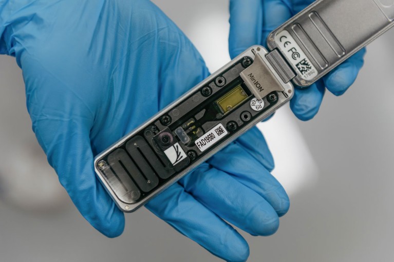
Two gloves hands holding a MinION portable and real time device for DNA and RNA sequencing

A nanopore sequencing device is typically used for sequencing DNA and RNA.Credit: Anthony Kwan/Bloomberg/Getty

With its fast analyses and ultra-long reads, nanopore sequencing has transformed genomics, transcriptomics and epigenomics. Now, thanks to advances in nanopore design and protein engineering, protein analysis using the technique might be catching up.

“All the pieces are there to start with to do single-molecule proteomics and identify proteins and their modifications using nanopores,” says chemical biologist Giovanni Maglia at the University of Groningen, the Netherlands. That’s not precisely sequencing, but it could help to work out which proteins are present. “There are many different ways you can identify proteins which doesn’t really require the exact identification of all 20 amino acids,” he says, referring to the usual number found in proteins.

In nanopore DNA sequencing, single-stranded DNA is driven through a protein pore by an electrical current. As a DNA residue traverses the pore, it disrupts the current to produce a characteristic signal that can be decoded into a sequence of DNA bases.

Proteins, however, are harder to crack. They cannot be consistently unfolded and moved by a voltage gradient because, unlike DNA, proteins don’t carry a uniform charge. They might also be adorned with post-translational modifications (PTMs) that alter the amino acids’ size and chemistry — and the signals that they produce. Still, researchers are making progress.

Water power

One way to push proteins through a pore is to make them hitch a ride on flowing water, like logs in a flume. Maglia and his team engineered a nanopore1 with charges positioned so that the pore could create an electro-osmotic flow that was strong enough to unfold a full-length protein and carry it through the pore. The team tested its design with a polypeptide containing negatively charged amino acids, including up to 19 in a row, says Maglia. This concentrated charge created a strong pull against the electric field, but the force of the moving water kept the protein moving in the right direction. “That was really amazing,” he says. “We really did not expect it would work so well.”

Chemists Hagan Bayley and Yujia Qing at the University of Oxford, UK, and their colleagues have also exploited electro-osmotic force, this time to distinguish between PTMs2. The team synthesized a long polypeptide with a central modification site. Addition of any of three distinct PTMs to that site changed how much the current through the pore was altered relative to the unmodified residues. The change was also characteristic of the modifying group. Initially, “we’re going for polypeptide modifications, because we think that’s where the important biology lies”, explains Qing.

And, because nanopore sequencing leaves the peptide chain intact, researchers can use it to determine which PTMs coexist in the same molecule — a detail that can be difficult to establish using proteomics methods, such as ‘bottom up’ mass spectrometry, because proteins are cut into small fragments. Bayley and Qing have used their method to scan artificial polypeptides longer than 1,000 amino acids, identifying and localizing PTMs deep in the sequence. “I think mass spec is fantastic and provides a lot of amazing information that we didn’t have 10 or 20 years ago, but what we’d like to do is make an inventory of the modifications in individual polypeptide chains,” Bayley says — that is, identifying individual protein isoforms, or ‘proteoforms’.

Molecular ratchets

Another approach to nanopore protein analysis uses molecular motors to ratchet a polypeptide through the pore one residue at a time. This can be done by attaching a polypeptide to a leader strand of DNA and using a DNA helicase enzyme to pull the molecule through. But that limits how much of the protein the method can read, says synthetic biologist Jeff Nivala at the University of Washington, Seattle. “As soon as the DNA motor would hit the protein strand, it would fall off.”

Nivala developed a different technique, using an enzyme called ClpX (see ‘Read and repeat’). In the cell, ClpX unfolds proteins for degradation; in Nivala’s method, it pulls proteins back through the pore. The protein to be sequenced is modified at either end. A negatively charged sequence at one end allows the electric field to drive the protein through the pore until it encounters a stably folded ‘blocking’ domain that is too large to pass through. ClpX then grabs that folded end and pulls the protein in the other direction, at which point the sequence is read. “Much like you would pull a rope hand over hand, the enzyme has these little hooks and it’s just dragging the protein back up through the pore,” Nivala says.
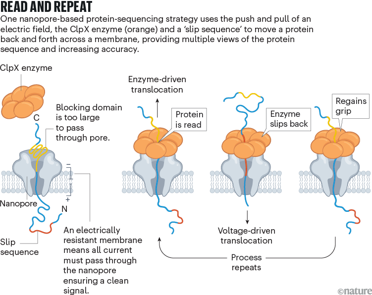
Read and repeat. Graphic showing a nanopore protein-sequencing strategy using the push and pull of an electric field through a membrane, enzyme and slip sequence.

Source: Ref. 3

Nivala’s approach has another advantage: when ClpX reaches the end of the protein, a special ‘slip sequence’ causes it to let go so that the current can pull the protein through the pore for a second time. As ClpX reels it back out again and again, the system gets multiple peeks at the same sequence, improving accuracy.

Last October3, Nivala and his colleagues showed that their method can read synthetic protein strands of hundreds of amino acids in length, as well as an 89-amino-acid piece of the protein titin. The read data not only allowed them to distinguish between sequences, but also provided unambiguous identification of amino acids in some contexts. Still, it can be difficult to deduce the amino-acid sequence of a completely unknown protein, because an amino acid’s electrical signature varies on the basis of both its surrounding sequence and its modifications. Nivala predicts that the method will have a ‘fingerprinting’ application, in which an unknown protein is matched to a database of reference nanopore signals. “We just need more data to be able to feed these machine-learning algorithms to make them robust to many different sequences,” he says.

Stefan Howorka, a chemical biologist at University College London, says that nanopore protein sequencing could boost a range of disciplines. But the technology isn’t quite ready for prime time. “A couple of very promising proof-of-concept papers have been published. That’s wonderful, but it’s not the end.” The accuracy of reads needs to improve, he says, and better methods will be needed to handle larger PTMs, such as bulky carbohydrate groups, that can impede the peptide’s movement through the pore.

How easy it will be to extend the technology to the proteome level is also unclear, he says, given the vast number and wide dynamic range of proteins in the cell. But he is optimistic. “Progress in the field is moving extremely fast.”

[ad_2]

Source Article Link

Categories
Life Style

DNA from ancient graves reveals the culture of a mysterious nomadic people

[ad_1]

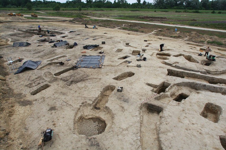
Excavation works conducted by the Eötvös Loránd University at the Avar-period (6th-9th century AD) cemetery of Rákóczifalva, Hungary, in 2006.

Scientists sampled genomic data from 279 graves at a cemetery in Rákóczifalva, Hungary, where people of the medieval Avar culture were buried.Credit: Institute of Archaeological Sciences, Eötvös Loránd University Múzeum, Budapest, Hungary

Most people know about the Huns, if only because of their infamous warrior-ruler Attila. But the Avars, another nomadic people who subsequently occupied roughly the same region of eastern and central Europe, have remained obscure despite having assembled a sprawling empire that lasted from the late sixth century to the early ninth century. Even archaeologists have struggled to piece together their history and culture, relying on spotty and potentially biased contemporaneous chronicles that, in many cases, were authored by the Avars’ adversaries.

A deep dive into 424 genomes collected from hundreds of Avar graves is filling in crucial gaps in this story, revealing a wealth of insights into the Avars’s social structure and culture1. “These people basically didn’t have a voice in history, and we are kind of looking into them this way — through their bodies,” says Zuzana Hofmanová, an archaeogeneticist at the Max Planck Institute for Evolutionary Anthropology in Leipzig, Germany, and one of the study’s lead authors.

The work was published today in Nature.

Nine generations

The researchers focused on four cemeteries in Hungary that were once at the heart of the khaganate, as the former Avar empire was known. Importantly, all four sites were fully excavated, giving the researchers access to DNA from every grave and enabling them to use genetic data to map relatedness for entire Avar communities.

This effort got an important boost from a computational method called ancIBD, which can connect even distant family members on the basis of their shared chromosomal sequences2. Co-lead author Johannes Krause, an archaeogeneticist at Max Planck, says that scientists have generally struggled to reassemble DNA-based family trees that extend past third-degree relatives, such as first cousins or great-grandparents. But by using tools such as ancIBD, Krause and colleagues were able to chart much more convoluted Avar family trees, including a massive nine-generation pedigree comprising 146 family members.

The data suggest that, after migrating to Europe, the Avars retained many cultural practices from their place of origin on the northeast Asian steppes3. For example, the Avars were very strict about avoiding inbreeding. There were no observed instances of marriage between relatives — even at the level of second cousins. Krasue says that was surprising, given that unions between first cousins were not unusual during much of European history. “It’s really remarkable that they can keep track over nine generations who is related to whom, and who can have children with whom,” he says.

On the other hand, there was also limited intermarriage with non-Avar neighbors: about 20% of the genomic sequences in the sampled Avar DNA could be traced to central European ancestry.

Gold figurine from the excavation at Rákóczifalva, Hungary.

A gold figurine excavated from an Avar burial site in Rákóczifalva, Hungary.Credit: Institute of Archaeological Sciences, Eötvös Loránd University Múzeum, Budapest, Hungary

The researchers recorded several examples of ‘levirate unions’, in which a widow married a male from the family of her deceased spouse, such as a brother. Such marital patterns were atypical in much of Europe, but were established features of Asian steppe-dwelling cultures, notes co-lead author Tivadar Vida, an archaeologist at Eötvös Loránd University in Budapest. “It was archaeologically very interesting to see the conservativism in the Avar society, lasting nine generations,” says Vida.

The Avars were also strictly patrilineal, with men acting as heads of family and daughters leaving their communities to join their husbands’ households. At the largest cemetery sampled, in the village of Rákóczifalva, Hungary, Hofmanová notes that there was only a single instance of both a mother and her adult daughter being interred.

Power play

The kinship data reveal what seems to be a shift in local political power that would have been difficult to detect with sparse DNA sampling. In the graves at Rákóczifalva, the researchers found that one male lineage predominated early in Avar history, but was displaced by a different Avar bloodline by the late seventh century. Intriguingly, archaeological evidence collected from those graves suggests that the subsequent family had different diets and burial rituals than did the displaced one, indicating that Avar culture shifted over time despite relatively modest levels of intermarriage with non-Avar individuals.

Carles Lalueza-Fox, a palaeogenomicist at the Institute of Evolutionary Biology in Barcelona, Spain, says that this work demonstrates the richness of the insights that can emerge when researchers have the opportunity and resources to broadly survey and analyse DNA at sites of historical interest. “Only this scale of analysis would allow you to obtain a reliable picture of kinship and social processes,” he says, adding that his group is now embracing a similar approach in their archaeogenomic research. “I think ancient genomics is moving toward this direction to obtain a more democratic and nuanced view of the past.”

[ad_2]

Source Article Link

Categories
Featured

Preparing for the DNA computation paradigm shift

[ad_1]

In “The Structure of Scientific Revolutions,” physicist and philosopher Thomas Kuhn introduced the concept of a paradigm shift, which he used to describe a fundamental change in the basic framework of thinking in natural sciences. Throughout history, however, such paradigm shifts have occurred not just in natural sciences but across the entire spectrum of human endeavor, providing solutions to problems that appeared to be insurmountable under the old paradigm.

The field of data storage and computation is a case in point. As the demand for creation, retention, and data computation only ever increased with time, the current computing paradigm requires enterprises to build data continuously centers the size of football fields and nuclear power plants to power them. Here, the lack of resources and capabilities to build these things quickly enough indefinitely is not as important as the fact that the current computing paradigm is not compatible with a scalable solution.

[ad_2]

Source Article Link

Categories
Featured

DNA storage is coming, it’s just a matter of when not if — SNIA quietly unveils first specifications for storing bytes in DNA medium, an important first step towards almost ultra-cheap, limitless storage

[ad_1]

Data will soon be able to be stored in DNA. French startup Biomemory has already shipped a DNA storage device to the general public. Now the DNA Data Storage Alliance, a technology affiliate of the Storage Networking Industry Association (SNIA), has unveiled the first specifications for storing vendor and CODEC information within a DNA data archive.

Unlike traditional storage mediums such as tape, HDD, and SSD, DNA lacks a fixed physical structure, necessitating a unique mechanism for reading or “booting up” a DNA archive.

[ad_2]

Source Article Link

Categories
Life Style

Memories are made by breaking DNA — and fixing it

[ad_1]

When a long-term memory forms, some brain cells experience a rush of electrical activity so strong that it snaps their DNA. Then, an inflammatory response kicks in, repairing this damage and helping to cement the memory, a study in mice shows.

The findings, published on 27 March in Nature1, are “extremely exciting”, says Li-Huei Tsai, a neurobiologist at the Massachusetts Institute of Technology in Cambridge who was not involved in the work. They contribute to the picture that forming memories is a “risky business”, she says. Normally, breaks in both strands of the double helix DNA molecule are associated with diseases including cancer. But in this case, the DNA damage-and-repair cycle offers one explanation for how memories might form and last.

It also suggests a tantalizing possibility: this cycle might be faulty in people with neurodegenerative diseases such as Alzheimer’s, causing a build-up of errors in a neuron’s DNA, says study co-author Jelena Radulovic, a neuroscientist at the Albert Einstein College of Medicine in New York City.

Inflammatory response

This isn’t the first time that DNA damage has been associated with memory. In 2021, Tsai and her colleagues showed that double-stranded DNA breaks are widespread in the brain, and linked them with learning2.

To better understand the part these DNA breaks play in memory formation, Radulovic and her colleagues trained mice to associate a small electrical shock with a new environment, so that when the animals were once again put into that environment, they would ‘remember’ the experience and show signs of fear, such as freezing in place. Then the researchers examined gene activity in neurons in a brain area key to memory — the hippocampus. They found that some genes responsible for inflammation were active in a set of neurons four days after training. Three weeks after training, the same genes were much less active.

The team pinpointed the cause of the inflammation: a protein called TLR9, which triggers an immune response to DNA fragments floating around the insides of cells. This inflammatory response is similar to one that immune cells use when they defend against genetic material from invading pathogens, Radulovic says. However, in this case, the nerve cells were responding not to invaders, but to their own DNA, the researchers found.

TLR9 was most active in a subset of hippocampal neurons in which DNA breaks resisted repair. In these cells, DNA repair machinery accumulated in an organelle called the centrosome, which is often associated with cell division and differentiation. However, mature neurons don’t divide, Radulovic says, so it is surprising to see centrosomes participating in DNA repair. She wonders whether memories form through a mechanism that is similar to how immune cells become attuned to foreign substances that they encounter. In other words, during damage-and-repair cycles, neurons might encode information about the memory-formation event that triggered the DNA breaks, she says.

When the researchers deleted the gene encoding the TLR9 protein from mice, the animals had trouble recalling long-term memories about their training: they froze much less often when placed into the environment where they had previously been shocked than did mice that had the gene intact. These findings suggest that “we are using our own DNA as a signalling system” to “retain information over a long time”, Radulovic says.

Fitting in

How the team’s findings fit with other discoveries about memory formation is still unclear. For instance, researchers have shown that a subset of hippocampal neurons known as an engram are key to memory formation3. These cells can be thought of as a physical trace of a single memory, and they express certain genes after a learning event. But the group of neurons in which Radulovic and her colleagues observed the memory-related inflammation are mostly different from the engram neurons, the authors say.

Tomás Ryan, an engram neuroscientist at Trinity College Dublin, says the study provides “the best evidence so far that DNA repair is important for memory”. But he questions whether the neurons encode something distinct from the engram — instead, he says, the DNA damage and repair could be a consequence of engram creation. “Forming an engram is a high-impact event; you have to do a lot of housekeeping after,” he says.

Tsai hopes that future research will address how the double-stranded DNA breaks happen and whether they occur in other brain regions, too.

Clara Ortega de San Luis, a neuroscientist who works with Ryan at Trinity College Dublin, says that these results bring much-needed attention to mechanisms of memory formation and persistence inside cells. “We know a lot about connectivity” between neurons “and neural plasticity, but not nearly as much about what happens inside neurons”, she says.

[ad_2]

Source Article Link