Categories
Life Style

Mini-colon and brain ‘organoids’ shed light on cancer and other diseases

[ad_1]

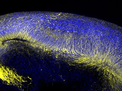
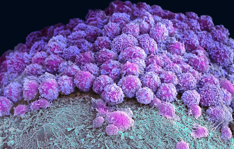
Coloured scanning electron micrograph (SEM) of a neural organoid.

Part of a brain organoid made using human stem cells (purple).Credit: Steve Gschmeissner/Science Photo Library

Researchers have hailed organoids — 3D clusters of cells that mimic aspects of human organs — as a potential way to test drugs and even eliminate some forms of animal experimentation. Now, in two studies published on 24 April in Nature1,2, biologists have developed gut and brain organoids that could improve understanding of colon cancer and help to develop treatments for a rare neurological disorder.

“In the last ten years, people spent a lot of time to develop and understand how to make organoids,” says Shuibing Chen, a chemical biologist at Weill Cornell Medical College in New York City. “But this is the time now to think more about how to use” the models.

Organoids — particularly those made from human stem cells — sometimes reveal things that animal models can’t, says Sergiu Pașca, a neuroscientist at Stanford University in California and a co-author of one of the studies1. Pașca’s group studies Timothy syndrome: a genetic disorder involving autism, neurological problems and heart conditions that affects only a few dozen people in the world. Timothy syndrome is caused by a single mutation in a gene called CACNA1C, which encodes a channel through which calcium ions enter cells including neurons.

Pașca says that there are no good animal models for Timothy syndrome because the underlying mutation doesn’t always cause the same symptoms in rodents. “It became very clear to us we’d need to find a way of testing in vivo,” he says.

Cultured cells

The researchers turned to brain organoids to recreate the disorder. They took stem cells from 3 people with the mutation that causes Timothy syndrome and cultured them for about 250 days, treating the cells with signalling molecules that encouraged them to turn into brain organoids containing every type of neuron found in the cerebral cortex, the outer layer of the brain. To create a more lifelike environment for the organoids, the team then injected these structures into the brains of rats, where the cells formed connections with the rodents’ own neurons. This made a system in which the researchers could test potential treatments for the disorder.

Human neurons have four different forms of this calcium channel, but only one of them is defective in Timothy syndrome. Getting rid of the mutated channel, the researchers suggest, would allow the other, healthy channels to take over.

To do this, they identified short pieces of nucleic acids called oligonucleotides that can stop cells producing the mutated form of the protein by interfering with genetic transcripts. Around two weeks after the researchers injected these oligonucleotides into the rats’ brains, most of the defective calcium channels in the organoids and the surrounding rat neurons had been replaced by other versions of the protein. The neurons in the organoids had also changed shape — from small, less complex forms similar to those in people with Timothy syndrome to larger, more complex shapes typical of healthy neurons. “To be honest, I didn’t think it would work so well,” Pașca says.

He adds that his group hope to test the therapy on people in clinical trials eventually, although they will first need to prove that the oligonucleotides are safe by testing them in non-human primates. The researchers think that the treatment would be effective for about three months, so people would need to receive frequent injections. The advantage, Pașca says, is that the biological effects of the treatment would be reversible and any side effects would be short-lived.

Miniature colon

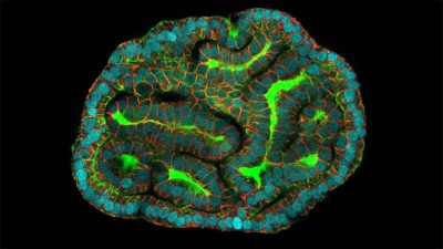
In a separate paper2, bioengineer Matthias Lütolf at the Swiss Federal Institute of Technology in Lausanne and his colleagues used mouse stem cells to model tissue from the other end of the body: the gut tissue that makes up the colon and rectum. Organoids tend to grow in tight balls, so the researchers grew the cells on a scaffold to recreate the tube structures seen in real gut tissue.

To make a model of colon cancer, they engineered the cells to contain light-sensitive proteins attached to cancer-causing genes. This allowed them to use a blue laser to switch on the genes and trigger the growth of tumours at specific sites in the organoid, then watch how the tumours changed over the course of weeks.
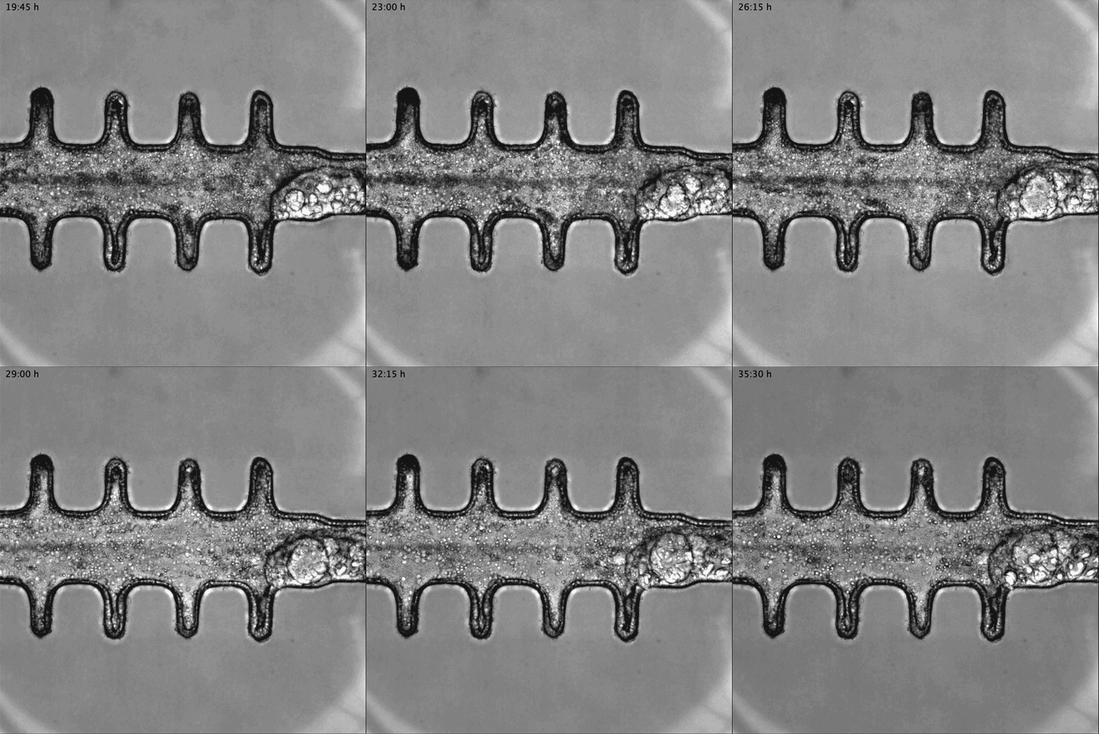
Animated sequence from a video showing mini-colon oncogenesis.

Researchers grew tumours inside a model of gut tissue created using mouse stem cells.Credit: Matthias P. Lutolf et al/Nature

When the researchers injected the cancerous cells into mice, the tumours looked similar to those seen in human colorectal cancer. The organoids accumulated fewer tumours when the researchers restricted calories in their medium, which also happens in people with colorectal cancer.

Lütolf was struck by the difference between the tumours triggered throughout the organoid. It would be difficult, he says, to use the organoids to screen a large number of new drugs, because of the differences and because they take a long time to produce. But he says organoids could be useful in investigating how drugs or immunotherapies kill tumours, and how factors such as a person’s environment and immune system affect the development and progression of colorectal cancer.

Lütolf and his group plan to manipulate the gut organoids to better reflect the human system, and hope that they could eventually replace animal models in some instances. Future organoid models, Lütolf says, could include bacteria that live in the gut or could be exposed to different levels of oxygen that mimic those available in various anatomical regions of the gut.

The two studies are “very well-designed”, Chen says. She adds that the organoid models seem particularly useful in showing the complexity of diseases such as Timothy syndrome and colorectal cancer, and how they progress over time. Now that researchers have found good ways to make organoids that model different organs, she says, the next steps will be learning how to scale up production for drug development, and making them more complex so that they reflect real human biology.

[ad_2]

Source Article Link

Categories
Life Style

Fungal diseases are spreading undetected

[ad_1]

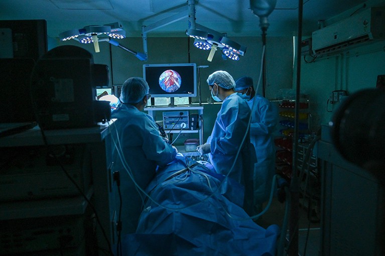
Two people wearing surgical gowns, face masks, and hair coverings, stand on either side of a patient (lying down in centre of image), performing surgery

Surgeons operate to remove ‘black fungus’ from a person’s lung.Credit: Prakash Singh/AFP via Getty

Lire en français

In mid-2021, a deadly fungal disease called mucormycosis began surging in India’s crowded hospitals. Often referred to as ‘black fungus’, and caused by various common mould species, mucormycosis typically invades structures in the face and brain. But during the Indian outbreak, thousands of people in hospital — most of them undergoing steroid treatment for severe COVID-19 — developed fungal infections that festered unseen in the lungs1. “We missed a lot of pulmonary mucormycosis during COVID,” says microbiologist Arunaloke Chakrabarti, director of the Doodhadhari Burfani Hospital and Research Institute in Haridwar, India. The expertise and tests needed to diagnose the condition were frequently unavailable, Chakrabarti says, and many people died before they could be treated.

India’s experience with mucormycosis reflects a broader diagnostic predicament: many low- and middle-income countries (LMICs) lack even the most basic tools for detecting the fungal diseases that kill an estimated 2.5 million people each year2. Misdiagnosis often leads to incorrect treatment, and, because people who are immunocompromised are particularly at risk, expanding use of steroids and other immune-suppressing drugs is contributing to the growth of vulnerable populations. People with HIV, cancer and respiratory diseases are especially at risk, particularly where poor sanitation and over-crowding aids the spread of fungal pathogens3.

As fungal disease rates rise steadily, efforts to shore up the ability of LMICs to diagnose the conditions have taken on a new urgency. In 2022, the World Health Organization (WHO) released its first-ever ranking of fungal health threats, with the aim of strengthening the global response to infections. The report emphasized that expanded access to diagnostics will help policymakers to better assess the burden of fungal diseases, so that resources and attention can be allocated more appropriately.

A global problem

Surveys of diagnostic capacity in LMICs over the past few years reveal a dire situation. Methods in many places are limited to conventional microscopy and fungal culture, both of which have shortcomings. Microscopy, for example, requires the expertise of people who can identify fungal pathogens by their appearance. Such specialists are typically unavailable in remote settings. And it can take up to one month to culture a sufficient quantity of fungal cells for microscopic analysis. That’s too long for people who are acutely ill, such as those “with fungal sepsis or fungal meningitis, who need a more immediate diagnosis”, says Marcio Louenco Rodrigues, a mycologist at the Oswaldo Cruz Foundation, a research institute in Curtiba, Brazil. Certain pathogens, including Pneumocystis jirovecii, which causes pneumonia, can’t be cultured at all.

In the absence of a diagnosis, physicians will often treat suspected infections on the basis of symptoms. But that approach comes with risks. Some fungal pathogens, including Mucorales, Histoplasma and Aspergillus cause lethal pulmonary infections that can be easily confused with tuberculosis, which is a bacterial disease4. Fungal infections don’t respond to antibiotics. And by the time people who are initially misdiagnosed with tuberculosis receive antifungal therapy, “many are already dying”, says Claudia Banda, an infectious-disease specialist at Cayetano Heredia University in Lima, Peru.

A key step towards boosting diagnostic capacity is to broaden access to simple, point-of-care assays that can be used cheaply in remote settings. “This is what developing countries really want,” says David Denning, a research clinician at the University of Manchester, UK. Denning was the founding president of the Global Action Fund for Fungal Infections (GAFFI), a non-governmental advocacy group headquartered in Geneva, Switzerland. GAFFI played a key part in selecting tests for fungal diseases for the WHO’s list of essential diagnostics, which was first released in 2018.

Topping that list are lateral flow assays that generate fast diagnostic results and cost less than US$5 each. These sorts of assay will be familiar to many as home-test kits for COVID-19. They detect microbial antigens or immune antibodies in blood and other fluids and have already been standardized for several fungal pathogens. For example, the WHO recommends screening people with HIV who have critically low white blood cell counts for cryptococcal antigen, which would indicate the presence of a harmful fungal infection that can cause meningitis5.

Lateral flow assays detect Cryptococcus with high accuracy. But in Africa — which is home to around 65% of people in the world with HIV — only around 25% of the population has access to the test, according to a survey funded by GAFFI3. And Latin American countries have similar shortages. In 2023, Banda and her colleagues surveyed Peru’s public hospitals, and found that only 13% of them could provide cryptococcal antigen testing (unpublished data).

A push to expand access

Uptake is thwarted by a lack of awareness of fungal infections, says Rodrigues, adding that for many fungal pathogens, there are no lateral flow tests. Cost is also a barrier, because although the price per test is low, rolling them out at scale would impose substantial burdens on already stretched health-care systems.

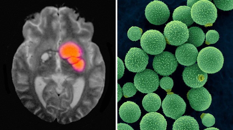
Image on left: black and white brain image with orange area to the right of centre. Image on Right: green spherical fungus particles of varying sizes, on dark background

A brain scan reveals cryptococcal meningitis, which can be caused by the fungus Cryptococcus neoformans (right).Credit: Cultura Creative RF/Alamy; Dennis Kunkel Microscopy/SPL

Routine use of the assay could save many lives — Cryptococcus kills nearly 150,000 people a year2. However, Banda points out that the main drug therapy for cryptococcal meningitis, amphotericin B, is also exceedingly toxic, “so we like to have some definitive evidence of the disease before we start treatment”.

Broadening diagnostics for other fungi that can cause serious illness, such as Aspergillus and Histoplasma, could also save lives. According to Denning, as many as 10% of people with suspected tuberculosis actually have chronic pulmonary aspergillosis. “So, all tuberculosis clinics around the world should be testing for it,” he says. Denning’s research on the global incidence of fungal diseases found that more than 2 million people each year develop invasive aspergillosis2, which occurs when the fungi spreads through the body. Histoplasmosis is also endemic in many LMICs, and if left untreated, mortality in people with HIV is 100%, Banda says.

Banda’s fungal-disease work in Peru, which is funded by the US Centers for Disease Control and Prevention, shows that histoplasmosis is endemic among people with HIV, cancer and other immune-suppressing conditions who live in and around the country’s coastal jungles. Bat droppings in these areas are a ready source of the fungus spores. Accurate estimates of the number of people affected are unavailable, however, owing to the lack of access to diagnostic testing, Banda says. She cites a similar need for access for chronic pulmonary aspergillosis and Candida auris — a hospital scourge that’s notoriously difficult to eliminate or control. The fungus C. auris, in particular, is an emerging global-health threat; some strains resist all antifungals and incur death rates as high as 70%. In Peru, the pathogen emerged several years ago, “and not all our microbiology labs are adequately prepared for it”, Banda says.

Advocates are calling for lateral flow assays to be developed for a broader array of fungal diseases. Along those lines, researchers at the University of Exeter, UK, are developing an assay specific to Rhizopus arrhizus6 — a fungal spore responsible for most human cases of mucormycosis, the disease that plagued India during the COVID-19 pandemic.

But lateral flow assays can’t solve the diagnostic shortages on their own. The tests are valuable as screening indicators that reveal whether a given pathogen is — or was — present in the body, but they don’t measure the extent of a fungal infection, or “how well a patient responds to treatment”, says Amir Seyedmousavi, a clinical microbiologist at the National Institutes of Health Clinical Center, in Bethesda, Maryland. Patient monitoring requires more advanced analytical techniques, Seyedmousavi says, such as polymerase chain reaction (PCR) sequencing and other molecular tests. The laboratory capacity needed for these technologies, however, is often unavailable in resource-poor areas.

Seyedmousavi chairs the Fungal Diagnostic Working Group of the International Society for Human and Animal Mycology. The group is working to standardize and improve affordable diagnostic tests — especially for LMICs. And it aims to create international networks for knowledge sharing and education. Chakrabarti, meanwhile, is working to establish diagnostic reference centres across India that offer a range of standardized diagnostics — not just at the hospitals at which they are based, but also at the clinics that serve as the first point of contact for health care. The reference centres provide training to boost laboratory workforces, Chakrabarti says, and staff there conduct epidemiology studies to map the distribution of fungal diseases throughout the country.

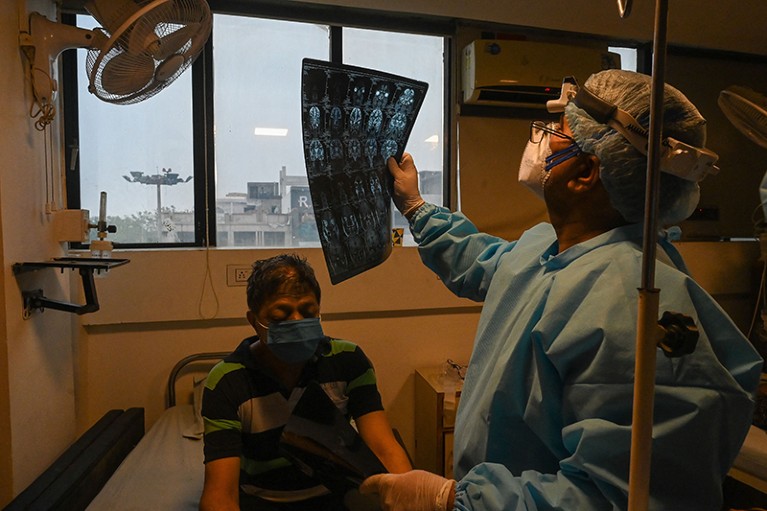
Person seated in room below window, wearing a face mask. To the right, person standing wearing PPE looking at MRI

During the peak of the COVID-19 pandemic, mucormycosis cases surged in India.Credit: Prakash Singh/AFP via Getty

GAFFI is sponsoring similar approaches in other parts of the world. In partnership with the non-profit organization the Asociacion de Salud Integral in Guatemala City, for instance, GAFFI sponsored a diagnostic demonstration that focused on a number of fungal diseases, including histoplasmosis and cryptococcosis. In 2023, GAFFI reported that mortality from fungal infections in people with HIV could be substantially reduced with readily available diagnostics and treatment.

Efforts to expand access to diagnostic tests face complex challenges. Because clinical awareness of fungal diseases is low in LMICs, physicians don’t request diagnostic tests often enough to create a market that could make the tests more readily available. In the absence of better information on prevalence — especially outside urban areas — health systems in low-income countries have failed to prioritize fungal diseases, Banda says, creating a vicious cycle of neglect.

Health-care financing poses extra hurdles. In some LMICs, people have to pay for fungal diagnostic tests out of their own pocket. “People don’t want to pay for a test that might be negative,” Denning says. “They just want the doctor to make a judgement about treatment. For the life-threatening fungal diseases — the ones that cause meningitis or sepsis or pneumonia — that can be tough.”

Denning argues that teaching hospitals in LMICs should have access to every test on the WHO’s list of essential fungal diagnostics, and that the tests should also be made freely available to people at the point of use. If hospitals don’t improve their capacity to detect and treat fungal diseases correctly, he warns, “then acquired microbial resistance and inappropriate use of all these antimicrobials just becomes rife”. Fortunately, diagnostic tests are steadily becoming cheaper, “so it should be possible to provide them at low-cost around the world,” Denning adds. “The big need is just to get them into everybody’s hands.”

[ad_2]

Source Article Link

Categories
Life Style

Ancient malaria genome from Roman skeleton hints at disease’s history

[ad_1]

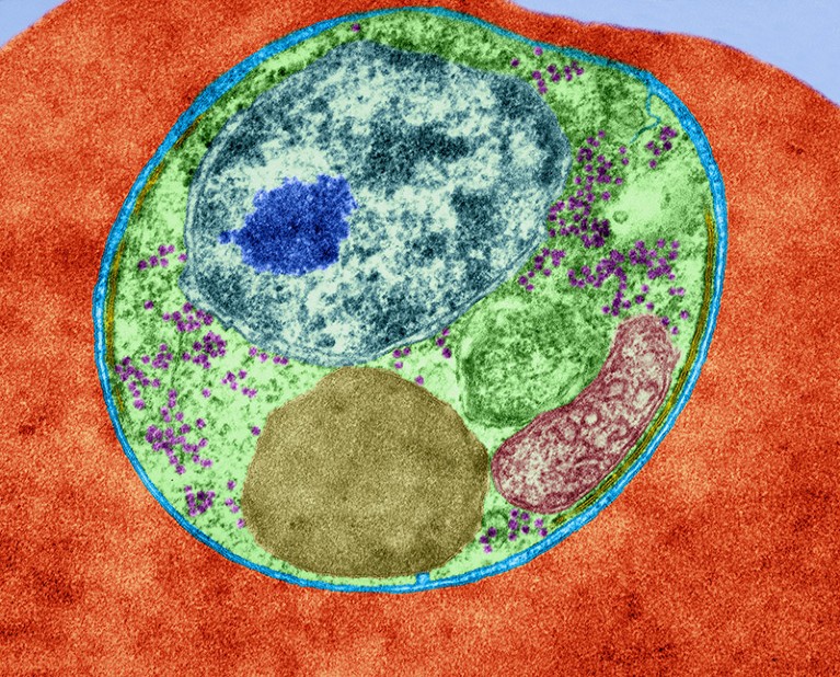
A coloured transmission electron micrograph showing a blue and green cell with several organelles inside a red cell.

The malaria parasite Plasmodium falciparum infecting a red blood cell.Credit: Dennis Kunkel Microscopy/Science Photo Library

Researchers have sequenced the mitochondrial genome of the deadliest form of malaria from an ancient Roman skeleton. They say the results could help to untangle the history of the disease in Europe.

It’s difficult to find signs of malaria in ancient human remains, and DNA from the malaria-causing parasite Plasmodium rarely shows up in them. As a result, there had never been a complete genomic sequence of the deadliest species, Plasmodium falciparum, from before the twentieth century — until now. “P. falciparum was eliminated in Europe a half century ago, and genetic data from European parasites — ancient or recent — has been an elusive piece in the puzzle of understanding how humans have moved parasites around the globe,” says Daniel Neafsey, who studies the genomics of malaria parasites and mosquito vectors at the Harvard T.H. Chan School of Public Health in Boston, Massachusetts.

Malaria has long been a leading cause of human deaths. “With the development of treatments such as quinine in the last hundreds of years, it seems clear [humans and malaria] are co-evolving,” says Carles Lalueza Fox, a palaeogenomicist at the Institute of Evolutionary Biology in Barcelona, Spain. “Discovering the genomes of the ancient, pre-quinine plasmodia will likely reveal information about how they have adapted to the different anti-malarial drugs.”

Ancient pathogen

There are five malaria-causing species of Plasmodium, which are thought to have arisen in Africa between 50,000 and 60,000 years ago, and then spread worldwide. Most researchers agree that they reached Europe at least 2,000 years ago, by the time of the Roman Empire.

Plasmodium falciparum “has significantly impacted human history and evolution”, says Neafsey. “So, that makes it particularly important to discover how long different societies have had to deal with [it], and how human migration and trade activities spread it.”

Researchers can glean valuable information about the origin, evolution and virulence of the parasite from DNA extracted from the ancient remains of infected people. But it is difficult to know where to look: it is not always obvious whether a person was infected with Plasmodium, and whether DNA can be recovered depends on how well it has been preserved.

In a preprint posted on the server bioRxiv1, a team of researchers led by a group at the University of Vienna identified the first complete mitochondrial genome sequence of P. falciparum from the bones of a Roman who lived in Italy in the second century ad, known as Velia-186.

Plasmodium falciparum had been detected in Velia-186 in a previous study2. The authors of the latest preprint extracted the parasite’s DNA from the body’s teeth, and were able to identify 5,458 pieces of unique genetic information that they combined to get a sequence covering 99.1% of the mitochondrial genome. They also used software to compare the genome with modern samples, and found that the Velia-186 sequence is closely related to a group of present-day strains found in India.

Carried by migration

The researchers say their findings support a hypothesis that P. falciparum spread to Europe from Asia around at least 2,000 years ago3. The Indian strains “were already present in Europe [then]; thus, a potential arrival with globalization episodes such as the Hellenistic period — when it is first described by Greeks — seems plausible”, says Lalueza Fox.

Neafsey says the work is a “technical tour de force” and an interesting addition to the limited field of ancient malaria genomics. But he adds that the results should be interpreted with caution because there are only a few samples, and points out that a genome sequence from DNA in the parasite’s cell nuclei, rather than its mitochondria, “might indicate a more complex story of parasite movement among ancient human populations”.

Lalueza Fox suggests exploring other potential sources of Plasmodium DNA, such as old bones, antique medical equipment and even mosquito specimens in museums. “The integration of genetic data from these heterogeneous sources will provide a nuanced view of this disease,” he says. “It would be interesting to see what lessons we can learn from the past on the strains and dispersals of this pathogen.”

[ad_2]

Source Article Link