Categories
Life Style

Why is exercise good for you? Scientists are finding answers in our cells

[ad_1]

When Bente Klarlund Pedersen wakes up in the morning, the first thing she does is pull on her trainers and go for a 5-kilometre run — and it’s not just about staying fit. “It’s when I think and solve problems without knowing it,” says Klarlund Pedersen, who specializes in internal medicine and infectious diseases at the University of Copenhagen. “It’s very important for my well-being.”

Whether it’s running or lifting weights, it’s no secret that exercise is good for your health. Research has found that briskly walking for 450 minutes each week is associated with living around 4.5 years longer than doing no leisure-time exercise1, and that engaging in regular physical activity can fortify the immune system and stave off chronic diseases, such as cancer, cardiovascular disease and type 2 diabetes. But, says Dafna Bar-Sagi, a cell biologist at New York University, the burning question is how does exercise deliver its health-boosting effects?

“We know that it is good, but there is still a huge gap in understanding what it is doing to cells,” says Bar-Sagi, who walks on a treadmill for 30 minutes, five days a week.

In the past decade, researchers have started to build a picture of the vast maze of cellular and molecular processes that are triggered throughout the body during — and even after — a workout. Some of these processes dial down inflammation, whereas others ramp up cellular repair and maintenance. Exercise also prompts cells to release signalling molecules that carry a frenzy of messages between organs and tissues: from muscle cells to the immune and cardiovascular systems, or from the liver to the brain.

But researchers are just beginning to work out the meaning of this cacophony of crosstalk, says Atul Shahaji Deshmukh, a molecular biologist at the University of Copenhagen. “Any single molecule doesn’t work alone in the system,” says Deshmukh, who enjoys mountain biking during the summer. “It’s an entire network that functions together.”

Exercise is also attracting attention from funders. The US National Institutes of Health (NIH), for instance, has invested US$170 million into a six-year study of people and rats that aims to create a comprehensive map of the molecules behind the effects of exercise, and how they change during and after a workout. The consortium behind the study has already published its first tranche of data from studies in rats, which explores how exercise induces changes across organs, tissues and gene expression, and how those changes differ between sexes24.

Building a sharper view of the molecular world of exercise could reveal therapeutic targets for drugs that mimic its effects — potentially offering the benefits of exercise in a pill. However, whether such drugs can simulate all the advantages of the real thing is controversial.

The work could also offer clues about which types of physical activity can benefit people with chronic illnesses, says Klarlund Pedersen. “We think you can prescribe exercise as you can prescribe a medicine,” she says.

Hard-wired for exercise

Exercise is a fundamental thread in the human evolutionary story. Although other primates evolved as fairly sedentary species, humans switched to a hunter-gatherer lifestyle that demanded walking long distances, carrying heavy loads of food and occasionally running from threats.

Those with better athletic prowess were better equipped to live longer lives, which made exercise a core part of human physiology, says Daniel Lieberman, a palaeoanthropologist at Harvard University in Cambridge, Massachusetts. The switch to a more active lifestyle led to changes in the human body: exercise burns up energy that would otherwise be stored as fat, which, in excess amounts, increases the risk of cardiovascular disease, type 2 diabetes and some cancers. The stress induced by running or pumping iron has the potential to damage cells, but it also kick-starts a cascade of cellular processes that work to reverse those effects. This can leave the body in better shape than it would be without exercise, says Lieberman.

Researchers have been exploring some of the biological changes that occur during exercise for more than a century. In 1910, pharmacologist Fred Ransom at the University of Cambridge, UK, discovered that skeletal muscle cells secrete lactic acid, which is created when the body breaks down glucose and turns it into fuel5. And in 1961, researchers speculated that skeletal muscle releases a substance that helps to regulate glucose during exercise6.

More clues were in store. In 1999, Klarlund Pedersen and her colleagues collected blood samples from runners before and after they took part in a marathon and found that several cytokines — a type of immune molecule — spiked immediately after exercise and that many remained elevated for up to 4 hours afterwards7. Among these cytokines were interleukin-6 (IL-6), a multifaceted protein that is a key player in the body’s defence response. The following year, Klarlund Pedersen and her colleagues discovered8 that IL-6 is secreted by contracting muscles during exercise, making it an ‘exerkine’ — the umbrella term for compounds produced in response to exercise.

A group of people doing tai chi outdoors with the Shanghai city skyline in the background.

Exercising regularly can strengthen the immune system and stave off disease.Credit: Mike Kemp/Getty

High levels of IL-6 can be beneficial or harmful, depending on how it is provoked. At rest, too much IL-6 has an inflammatory effect and is linked to obesity and insulin resistance, a hallmark of type 2 diabetes, says Klarlund Pedersen. But when exercising, the molecule activates its more calming family members, such as IL-10 and IL-1ra, which tone down inflammation and its harmful effects. “With each bout of exercise, you provoke an anti-inflammatory response,” says Klarlund Pedersen. Although some physical activity is better than none, high-intensity, long-duration exercise that engages large muscles — such as running or cycling — will crank up IL-6 production, adds Klarlund Pedersen.

Exercise is a balancing act in other ways, too. Physical activity produces cellular stress, and certain molecules counterbalance this damaging effect. When mitochondria — the powerhouses that supply energy in cells — ramp up production during exercise, they also produce more by-products called reactive oxygen species (ROS), which, in excessive amounts, can damage proteins, lipids and DNA. But these ROS also kick-start a horde of protective processes during exercise, offsetting their more toxic effects and fortifying cellular defences.

Among the molecular stars in this maintenance and repair arsenal are the proteins PGC-1α, which regulates important skeletal muscle genes, and NRF2, which activates genes that encode protective antioxidant enzymes. During exercise, the body has learnt to benefit from a fundamentally stressful process. “If stress doesn’t kill you it makes you stronger,” says Ye Tian, a geneticist at the Institute of Genetics and Developmental Biology, Chinese Academy of Sciences, Beijing.

Exerkines everywhere

Since IL-6 ushered in the exerkine era, the explosion of multiomics — an approach that combines various biological data sets, such as the proteome and metabolome — has allowed researchers to go beyond chasing single molecules. They can now begin untangling the convoluted molecular web that lies behind exercise, and how it interacts with different systems across the body, says Michael Snyder, a geneticist at Stanford University in California, who recently switched from running to weightlifting. “We need to understand how these all work together, because [humans] are a homeostatic machine that needs to be properly tuned,” he says.

In 2020, Snyder and his colleagues took blood samples from 36 people aged between 40 and 75 years old before, during and at various time intervals after the volunteers ran on a treadmill. The team used multiomic profiling to measure more than 17,000 molecules, more than half of which showed significant changes after exercise9. They also found that exercise triggered an elaborate ‘choreography’ of biological processes such as energy metabolism, oxidative stress and inflammation. Creating a catalogue of exercise molecules is an important first step in understanding their effects on the body, says Snyder.
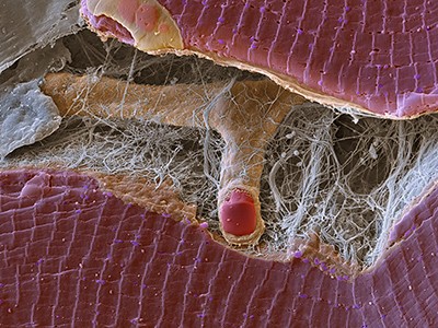
Other studies have probed how exercise affects cell types. A 2022 study in mice led by Jonathan Long, a pathologist at Stanford University, identified more than 200 types of protein that were expressed differently by 21 cell types in response to exercise10. The researchers were expecting to find that cells in the liver, muscle and bone would be most sensitive to exercise, but to their surprise, they found that a much more widespread type of cell, one that appears in many tissues and organs, showed the biggest changes in the proteins that it cranked out or turned down. The findings suggest that more cell types shift gears during a workout than was previously thought, although what these changes mean for the body is still an open question, says Long.

The findings also showed that after exercise, the mice’s liver cells squeezed out several types of carboxylesterase enzyme, which are known to ramp up metabolism. When Long and his colleagues genetically tweaked mice so that their livers expressed elevated levels of these metabolism-enhancing enzymes, and then fed them a diet of fatty foods, the mice didn’t gain weight. They also had increased endurance when they ran on a treadmill. “The improvement in exercise performance by these secreted carboxylesterases was not known before,” says Long, whose weekly exercise regime involves swimming and lifting weights. He adds that if the enzymes could be produced in the right quantities and purity, they could possibly be used as exercise-mimicking compounds.

During a workout, distant organs and tissues communicate with each other through molecular signals. Along with exerkines, extracellular vesicles (EVs) — nanosized, bubble-shaped structures that carry biological material — could be one of the mechanisms behind organ and tissue crosstalk, says Mark Febbraio, a former triathlete who is now an exercise physiologist at Monash University in Melbourne, Australia. In 2018, Febbraio and his team inserted tubes into the femoral arteries of 11 healthy men and drew blood before and after they rode an exercise bike at an increasing pace for an hour. During and after exercise, but not at rest, they found a spike in the levels of more than 300 types of protein that compose or are carried by EVs11.

When the team then collected EVs from mice that had run on a treadmill and injected them into another group of healthy mice, most of the EVs ended up in liver cells. In a separate mouse study that is yet to be published, Febbraio and his colleagues found hints that the contents of these liver-bound EVs can arrest a type of liver disease. A big question is whether EVs also deposit genetic material into different cells, and if so, what that means for the body. “We still don’t know a great deal,” he says.

Exercise as medicine

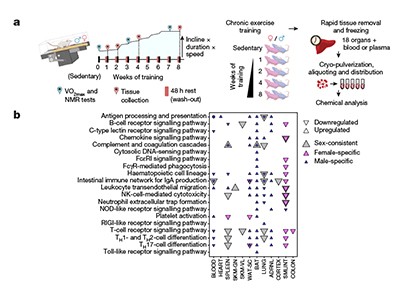
Larger efforts are under way to build a detailed molecular snapshot of how exercise exerts its health-boosting effects across tissues and organs. In 2016, the NIH established the Molecular Transducers of Physical Activity Consortium (MoTrPAC), a six-year study on around 2,600 people and more than 800 rats that aims to generate a molecular map of exercise. The effort — one of the largest studies on physical activity — is teasing apart the effects of aerobic and endurance exercise on multiple tissue types across different ages and fitness levels.

The first data set is from rats that completed one to eight weeks of treadmill training, and had blood and tissue samples collected at the end. The researchers pinpointed thousands of molecular changes throughout the rats’ bodies, many of which could have a protective effect on health, such as dialling down inflammatory bowel disease and tissue injury2. A separate study3 found that the effects of endurance training differed across sexes: markers associated with the breakdown of fat increased in male fat tissue, driving fat loss, whereas female fat tissue showed an increase in markers related to fat-cell maintenance and insulin signalling, which might protect against cardiometabolic diseases. A third study4 found that exercise alters the expression of genes linked to diseases such as asthma, and could help to trigger similar adaptive responses.

A big goal is to uncover why exercise has such varied effects on people of different sexes, ages and ethnic backgrounds, says Snyder, who is a member of the MoTrPAC team. “It’s very obvious that some people benefit better than others,” he says.

Researchers hope that the reams of molecular data will eventually help clinicians to develop tailored exercise prescriptions for people with chronic diseases, says MoTrPAC team member Bret Goodpaster, an exercise physiologist at the University of Pittsburgh in Pennsylvania. Farther down the track, such insights could be used to develop therapeutics that mimic some of the beneficial effects of exercise in people who are too ill to work out, he says. “That’s not to say that we will have exercise in a pill, but there are certain aspects of exercise that could be druggable,” says Goodpaster, who has taken part in triathlons, marathons and cycling races.

Several teams are already in the early stages of developing exercise-mimicking therapeutics. In March 2023, a team led by Thomas Burris, a pharmacologist at the University of Florida in Gainesville, identified a compound that targets proteins called oestrogen-related receptors, which are known to trigger key metabolic pathways in energy-intensive tissues, such as heart and skeletal muscle, particularly during exercise12. When the researchers administered the compound — called SLU-PP-332 — to mice, they found that the treated rodents were able to run 70% longer and 45% farther than untreated mice. Six months later, a separate study, also led by Burris, found that obese mice treated with the drug lost weight and gained less fat than those that didn’t receive the treatment — even though their diet was the same and they didn’t exercise any more than usual13.

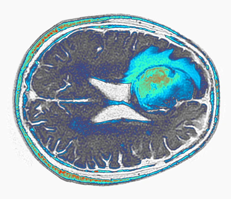
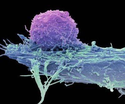
There is already evidence that exercise itself acts like medicine. In 2022, Bar-Sagi and her colleagues found that mice with pancreatic cancer had elevated levels of CD8 T cells — which destroy cancerous and virus-infected cells — when they did 30 minutes of aerobic exercise for 5 days a week14. These killer cells express a receptor for IL-15, another exerkine released by muscles during exercise. The researchers found that when CD8 T cells bind to IL-15, they unleash a more powerful immune response on tumours in the pancreas. This effect prolonged survival of mice with tumours by around 40%, compared with that of control mice. The findings held up when Bar-Sagi and her team analysed tumour tissue taken from people with pancreatic cancer. Those who did 60 minutes of aerobic and strength training each week had more CD8 T cells, and were twice as likely to survive for up to 5 years, than were people in the control group.

Although exercising more is a no-brainer for improving health, around 25% of adults globally do not meet the World Health Organization’s recommended levels of exercise each week: 150–300 minutes or more of moderate-intensity exercise, such as a brisk walk; or 75–150 minutes of vigorous-intensity exercise, such as running. David James, an exercise physiologist at the University of Sydney in Australia, who rides his bike to work each day, says that understanding the inner workings of exercise could help to develop clearer public-health messages about why physical activity is important and how it can offset the risk of getting chronic diseases. “That’s a powerful message,” says James.

[ad_2]

Source Article Link

Categories
Life Style

85 million cells — and counting — at your fingertips

[ad_1]

When it comes to single-cell gene-expression data, biologists face an embarrassment of riches. There are thousands of data sets to choose from. Unfortunately, those data sets have not all been processed in the same way; they might use different names for similar or identical cells or tissues; and they are scattered across the Internet — or available only on request.

Using any one data set is relatively straightforward. But collecting, curating and integrating the data to draw conclusions across experiments, is — in the words of bioinformatician Timothy Triche Jr at the Van Andel Institute in Grand Rapids, Michigan — “a huge pain in the butt”.

In one 2023 study1, for instance, computational biologist Christina Theodoris at Gladstone Institutes in San Francisco, California, described a deep-learning model called Geneformer. Building on some 30 million single-cell transcriptomic data sets that Theodoris manually aggregated in 2021, Geneformer allows researchers to predict the impact of gene perturbations in cell types or genes it has never seen. But because the data were scattered across 18 public databases and multiple independent laboratories, she says, “it took me two months to collect all that data and process it”.

A vast resource

Today, the same effort would take only minutes, she says, thanks to a new resource from the Chan Zuckerberg Initiative (CZI) in Redwood City, California. Chan Zuckerberg CELL by GENE Discover (CZ CELLxGENE) is a collection of free and open-source tools for finding, querying, analysing, downloading and publishing single-cell data. As of April, it includes some 85 million single cells and 1,317 data sets covering 844 cell types, curated and uniformly processed by a team of 25 or so engineers, data curators and other staff, according to Patricia Brennan, vice-president of science technology at CZI. Most of the data represent single-cell RNA sequencing information from healthy human tissues, but non-human and cell-line data, as well as molecular-profiling data obtained using spatial transcriptomic methods, are also available. All of these data are stored in a common format, using a standard set of cell types and metadata.

A laptop screen showing a UMAP plot within the Chan Zuckerberg CELL by GENE platform

The CZ CELLxGENE tool helps researchers to visualize gene-expression data.Credit: Chan Zuckerberg Initiative

Users can find and explore the non-spatial data through the CZ CELLxGENE data portal, or access it using the R or Python programming languages through an application-programming interface called Census. (Spatial data should be added later this year, a spokesperson for CZI says.) Meera Prasad, a graduate student at the California Institute of Technology in Pasadena, is using CZ CELLxGENE to characterize the microenvironment across 9 million healthy and cancerous mammary cells representing some 150 cell types. By integrating those data with her lab’s spatial data, Prasad hopes to better replicate the tumour microenvironment, and also to identify genes that are related to the structural changes associated with cancer.

CZ CELLxGENE enables two key applications, says Jonah Cool, a science programme officer at CZI. Most obviously, researchers can ask questions across a vast amount of data that they and others have collected. Triche, for instance, has plumbed some 12 million mouse cells to study the influence of sex chromosomes on the biology of immune cells. “That’s approximately 11-and-a-half million more cells than we would typically run in a single-cell experiment,” he says. Repeating those analyses in-house would be a waste of money, but leveraging data that others have processed can be tedious. By ‘harmonizing’ these data sets and putting them in one place, CZ CELLxGENE removes many of what Triche calls “schlep steps”. “People underestimate the degree to which the impact of this data is amplified by making it usable for anybody who wants to,” he says.

The other application is in artificial intelligence. Researchers can use CZ CELLxGENE to build and train computational models that can predict, for instance, the identity of a cell or the impact of specific perturbations.

Model modularity

Users can select any of five such models, including Geneformer, and refine or apply them to their own data. They can also download ‘embeddings’ — compressed numerical representations of transcriptional data — from any of them, allowing users to ‘project’ their data and CZ CELLxGENE data into a common space. That, says Cool, means researchers can ask questions such as what cells are similar to a researcher’s cells, or which conditions induce changes in those cells.

Computer scientist Jure Leskovec at Stanford University in California, used his Universal Cell Embeddings model2, which he trained on CZ CELLxGENE data, to identify rare mouse kidney cells known as Norn cells. By then applying this ‘classifier’ to a larger data set of 36 million cells, he found that Norn cells were also present in the heart, lung and gonads. “This generalizability is the key capability of these models,” he says.

CZ CELLxGENE is not the only resource that aggregates and simplifies single-cell data analysis. The Human Cell Atlas, for instance, has its own data portal. And both the University of California, Santa Cruz, and the Broad Institute of MIT and Harvard in Cambridge, Massachusetts, among others, host tools for analysing select single-cell data sets online.

In March, Lior Pachter, a computational biologist at the California Institute of Technology, and his team described their Commons Cell Atlas infrastructure3,4 that stores and uniformly processes raw sequence data across data sets. (By contrast, CZ CELLxGENE retains data as ‘gene-count matrices’, although links to the original sequence data are also maintained, a spokesperson says.) These sequence data can be reanalysed as gene annotations change, Pachter notes, and his team exploited that to study gene-splice isoforms in human testis. “It’s really powerful and useful to be able to go back and rebuild the atlas again and again and again,” he says.

In September 2023, CZI announced that it would build a computing cluster of 1,000 graphical processing units (GPUs), which can rapidly accelerate or scale up model development.

This is helpful to researchers because most labs doing single-cell research, Cool explains, have access to maybe a handful of GPUs, therefore limiting the complexity of the models that they can build and lengthening experiments. Using the new cluster, Cool says, researchers can begin to build more sophisticated — and accurate — models. The cluster is expected to be “up and running by June”, a spokesperson says.

[ad_2]

Source Article Link

Categories
Life Style

Protein in embryo cells might be a reason for right- or left-handedness

[ad_1]

Hello Nature readers, would you like to get this Briefing in your inbox free every day? Sign up here.

A young left handed child draws with a crayon on an easel.

Credit: incamerastock/Alamy

Left-handed people are almost three times more likely to have rare variants in the genes for tubulins, proteins that build cells’ internal skeletons. Tubulins assemble into long filaments called microtubules, which control the shapes and movements of cells. Microtubules could influence handedness because they form hair-like protrusions in cell membranes that can direct fluid flows in an asymmetric way during embryonic development.

Nature | 4 min read

Reference: Nature Communications paper

Super-Earth LHS 3844b is the first exoplanet to show convincing signs of tidal synchronization, meaning one of its hemispheres is permanently illuminated by its star and the other is in permanent darkness. The planet is relatively cool, indicating that it lacks the tidal heating non-locked planets experience. It’s unclear whether tidally locked planets could be habitable. These worlds “don’t have tides, or seasons or day–night cycles”, says astronomer and study co-author Nicolas Cowan. “Could you get the same kind of diversity and complexity of life evolving? I have no idea.”

Nature | 4 min read

Reference: The Astrophysical Journal paper

Biotechnology company LyGenesis has injected donor liver cells into a lymph node of a person with liver failure for the first time. The idea is that, within months, the donor cells will grow into a blood-filtering ‘miniature liver’. “It’s a very bold and incredibly innovative idea,” says liver-regeneration specialist Valerie Gouon-Evans. The treatment, which has been trialled in mice, dogs and pigs, might not relieve all of the complications of end-stage liver disease — but could provide a stopgap until a donor organ becomes available or make people healthy enough to undergo a transplant.

Nature | 5 min read

Nearly 20% of almost 600 image-containing papers that scientists compiled for a systematic review had suspicious images, including those that had been duplicated, stretched or rotated. Out of the 132 studies the researchers included in their final review, which was about a test to identify depression-related symptoms in rats, 10 contained potentially doctored images. Analysing these 10 alone assessed the test as 50% more effective than did the remaining 122 studies. This “clearly highlights [that falsified images] are impacting our consolidated knowledge base”, says systematic-review methodologist Alexandra Bannach-Brown.

Nature | 5 min read

Reference: bioRxiv preprint (not peer reviewed)

Features & opinion

When microbes’ ability to develop is pitted against their ability to make pharmaceuticals or biofuels, “the cells are going to choose to grow every time”, says synthetic biologist Brian Pfleger. To sidestep this unwinnable metabolic-resource war, researchers are introducing new biosynthesis pathways that can run alongside natural processes. For this, they have zeroed in on cofactors, small molecules that help enzymes do their work. Eventually, synthetic cofactors paired with the enzymes that use them could allow cells to churn out compounds more efficiently or even make those that rarely occur in nature.

Nature | 11 min read

With Twitter on the wane among many scientists — both as a place to post and a source of social-media data — some are turning to Reddit. Access to Reddit data is free for non-commercial researchers and academics. And its communities offer a space to network and chat, with an ‘upvote and downvote’ system helping good content rise to the top. Nature offers some context and advice for those looking to take the plunge.

Nature | 10 min read

Cancer cells make proteins found nowhere else in the body. Vaccines could teach the body’s immune system to recognise these proteins and destroy the cancer cells. The most powerful vaccines are created from the specific proteins extracted from a patient’s tumour and, in some cases, use the patient’s own immune cells. Researchers are also working on doing this vaccination process entirely within the body: first, drugs activate the immune system, then radiotherapy kills cancer cells, releasing the cancer proteins for the switched-on immune cells to find.

Nature | 4 min video

This article is part of Nature Outline: Cancer vaccines, an editorially independent supplement produced with the financial support from Moderna.

Where I work

Muh Aris Marfai measures the ground level compared to level zero of the sea (average sea level) using a geodetic digital level, at a tide gauge station at the Sunda Kelapa port of Jakarta, Indonesia.

Muh Aris Marfai is head of the Geospatial Information Agency of Indonesia in Bogor and a geography researcher at the Gadjah Mada University in Yogyakarta.Credit: Gaia Squarci for Nature

Geographer Muh Aris Marfai collects reference data for Indonesia’s coastal areas to prepare for the impacts of climate change. “Because so much of the country is surrounded by water, it’s important to pay attention to coastal areas,” he says. “Many coastal cities, including Jakarta, are experiencing subsidence owing to geological processes and coastal dynamics.” (Nature | 3 min read)

Quote of the day

Psychologist Jonathan Haidt’s blockbuster new book The Anxious Generation suggests that digital technologies are rewiring our children’s brains and causing an epidemic of mental illness — but its claims are not backed up by science, writes psychologist Candice Odgers. (Nature | 6 min read)

Today I’m enjoying the fish doorbell — a charming solution to alert the lock keepers of Utrech’s boat canal to let through migrating fish. A webcam allows watchers to ‘ring the doorbell’ for the fish, sending a photo to ecologists who signal that it’s time to clear the underwater traffic jam. The doorbell has struck a chord with nature-lovers, meaning the 950-ish slots for aspiring doorbell-ringers are often full. And even if you do get access, you might wait days to spot a fish in the murky waters. But all the enthusiasm is good news for ecologist Mark van Heukelum, who created the gadget. “We ‘only’ get a thousand pictures of every fish that appears for the camera,” he jokes on the fish-doorbell website.

Thanks for reading,

Flora Graham, senior editor, Nature Briefing

With contributions by Katrina Krämer

Want more? Sign up to our other free Nature Briefing newsletters:

Nature Briefing: Anthropocene — climate change, biodiversity, sustainability and geoengineering

Nature Briefing: AI & Robotics — 100% written by humans, of course

Nature Briefing: Cancer — a weekly newsletter written with cancer researchers in mind

Nature Briefing: Translational Research covers biotechnology, drug discovery and pharma

[ad_2]

Source Article Link

Categories
Life Style

Right- or left-handed? Protein in embryo cells might help decide

[ad_1]

A young left handed child draws with a crayon on an easel.

Dozens of genetic factors have been associated with left-handedness, which occurs in roughly one in ever ten people.Credit: incamerastock/Alamy

To what extent do genes determine how you pick up your morning cup of coffee? Researchers examined rare genetic variants from a database of more than 350,000 individuals’ genetic data to hunt for clues for what influences handedness in humans. Their findings implicate tubulins — proteins that build cells’ internal skeletons.

The results, published on 2 April in Nature Communications1, were obtained specifically at protein-coding parts of the DNA, and add to previous studies that linked genetic variations with handedness .

“This is an important and significant study” that supports tubulins’ involvement in determining the left–right brain asymmetry, says Sebastian Ocklenburg, a neuroscientist at the Medical School Hamburg in Germany.

During the embryonic stage of human development, the left and right brain hemispheres get wired differently, which in part determines innate behaviours, such as where we lean when we hug someone, on which side of our mouth we tend to chew our food and, most prominently, which hand is our dominant one. This turns out to be the left hand for around 10% of the human population.

Because most people have a clear preference for one hand over the other, finding genes linked to handedness can provide clues for the genetic basis of the brain’s left–right asymmetry.

Previous studies looking at genome-wide data from UK Biobank2 found 48 common genetic variants associated with left-handedness, which were mostly in non-coding regions of the DNA. These included sections that could control the expression of genes related to tubulins. These proteins assemble into long, tube-like filaments called microtubules, which control the shapes and movements of cells.

But Clyde Francks, a geneticist and neuroscientist at the Max Planck Institute for Psycholinguistics in Nijmegen, the Netherlands, and his team looked for genetic variants in protein-coding sequences. Their analysis of 313,271 right-handed and 38,043 left-handed individuals’ genetic data, from the UK Biobank, uncovered variants in a tubulin gene, dubbed TUBB4B, which were 2.7 times more common in left-handed people than in right-handers.

Microtubules could influence handedness because they form cilia — hair-like protrusions in cell membranes — which can direct fluid flows in an asymmetric way during development.

In spite of affecting only a small proportion of the people in this considerable data set, rare variants “can give clues to developmental mechanisms of brain asymmetry in everyone”, Francks says. He adds that these findings pave the way for future work to determine how microtubules, which themselves have a molecular ‘handedness’, can give an “asymmetric twist” to early brain development.

[ad_2]

Source Article Link

Categories
Life Style

These ‘movies’ of proteins in action are revealing the hidden biology of cells

[ad_1]

Since the 1950s, scientists have had a pretty good idea of how muscles work. The protein at the centre of the action is myosin, a molecular motor that ratchets itself along rope-like strands of actin proteins — grasping, pulling, releasing and grasping again — to make muscle cells contract.

The basics were first explained in a pair of landmark papers in Nature1,2, and they have been confirmed and elaborated on by detailed molecular maps of myosin and its partners. Researchers think that myosin generates force by cocking back the long lever-like arm that is attached to the motor portion of the protein.

The only hitch is that scientists had never seen this fleeting pre-stroke state — until now.

In a preprint published in January3, researchers used a cutting-edge structural biology technique to record this moment, which lasts just milliseconds in living cells.

“It’s one of the things in the textbook you sort of gloss over,” says Stephen Muench, a structural biologist at the University of Leeds, UK, who co-led the study. “These are experiments that people wanted to do 40 years ago, but they just never had the technology.”

That technology — called time-resolved cryo-electron microscopy (cryo-EM) — now has structural biologists thinking like cinematographers, turning still snapshots of life’s molecular machinery into motion pictures that reveal how it works.

Muench and his colleagues’ myosin movie isn’t feature-length; it consists of just two frames showing different stages of the molecular motion. Yet it confirmed a decades-old theory and settled debates over the order of the steps in myosin’s choreography. Other researchers are focusing their new-found director’s eye on understanding cell-signalling systems, including those underlying opioid overdoses, the gene-editing juggernaut CRISPR–Cas9 and other molecular machines that have been mostly studied with highly detailed, yet static structural maps.

An animated gif showing a 3D molecular structures of a myosin molecule in two states using a lever arm to pull on an actin fillament

Researchers have been able to capture images of individual myosin proteins as they pull on an actin filament during muscle contraction, confirming key details of the motion. First, myosin becomes cocked or primed, then it attaches to actin and its lever arm swings in a power stroke that slides the filament by about 34 nanometres.Credit: Sean McMillan

“The big picture is to move away, as much as possible, from this single, static snapshot,” says Georgios Skiniotis, a structural biologist at Stanford University in California, whose team used the technique to record the activation of a type of cell-signalling molecule called a G-protein-coupled receptor (GPCR)4. “I want the movie.”

Freeze frame

To underscore the power of cryo-EM, Skiniotis and others like to draw a comparison with one of the first motion pictures ever made. In the 1870s, photographer Eadweard Muybridge used high-speed photography technology, which was cutting edge at the time, to capture a series of still images of a galloping horse. They showed, for the first time, that all four of the animal’s hooves leave the ground at once — something that the human eye could not distinguish.

Similar insights, Skiniotis says, will come from applying the same idea to protein structures. “I want to get a dynamic picture.”

The ability to map proteins and other biomolecules down to the location of individual atoms has transformed biology, underpinning advances in gene editing, drug discovery and revolutionary artificial-intelligence tools such as AlphaFold, which can predict protein structures. But the mostly static images delivered by X-ray crystallography and cryo-EM, the two technologies responsible for the lion’s share of determined protein structures, belie the dynamic nature of life’s molecules.

“Biomolecules are not made up of rocks,” says Sonya Hanson, a computational biophysicist at the Flatiron Institute in New York City. They exist in water and are constantly in motion. “They’re more like jelly,” adds Muench.

Biologists often say that “structure determines function”, but that’s not quite right, says Ulrich Lorenz, a molecular physicist at the Swiss Federal Institute of Technology in Lausanne (EPFL). The protein poses captured by most structural studies are energetically stable ‘equilibrium’ states that provide limited clues to the short-lived, unstable confirmations that are key to chemical reactions and other functions performed by molecular machines. “Structure allows you to infer function, but only incompletely and imperfectly, and you’re missing all of the details,” says Lorenz.

Cryo-EM is a great way to get at the details, but capturing these fleeting states requires careful preparation. Protein samples are pipetted onto a grid and then flash frozen with liquid ethane. They are then imaged using powerful electron beams that record snapshots of individual molecules (sophisticated software classifies and morphs these pictures into structural maps). The samples swim in water before being frozen, so any chemical reaction that can happen in a test tube can, in theory, be frozen in place on a cryo-EM grid — if researchers can catch it quickly enough.

That’s one of the first big challenges says Joachim Frank, a structural biologist at Columbia University in New York City who shared the 2017 Nobel Prize in Chemistry for his work on cryo-EM. “Even for very dexterous people, it takes a few seconds.” In that time, any chemical reactions — and the intermediate structures that mediate the reactions — might be long gone before freezing. “This is the gap we want to fill,” says Frank.

Caught in translation

Frank’s team has attempted to solve this problem using a microfluidic chip. The device quickly mixes two protein solutions, allows them to react for a specified time period and then delivers reaction droplets onto a cryo-EM grid that is instantly frozen.

This year, Frank’s team used their device to study a bacterial enzyme that rescues ribosomes, the cell’s protein-making factories, if they stall in response to antibiotics or other stresses. The enzyme, called HflX, helps to recycle stuck ribosomes by popping their two subunits apart.

Frank’s team captured three images of HflX bound to the ribosome, over a span of 140 milliseconds, which show how it splits the ribosome like someone carefully removing the shell from an oyster. The enzyme breaks a dozen or so molecular bridges that hold a ribosome’s two subunits together, one by one, until just two are left and the ribosome pops open5. “The most surprising thing to me is that it’s a very orderly process,” Frank says. “You would think the ribosome is being split and that’s it.”

Muench and his colleagues, including Charlie Scarff, a structural biologist at the University of Leeds, and Howard White, a kineticist at Eastern Virginia Medical School in Norfolk, Virginia also used a microfluidic chip to make their myosin movie by quickly mixing myosin and actin3.

But the molecular motor is so fast that, to slow things down ever further, they used a mutated version of myosin that operates about ten times slower than normal. This allowed the team to determine two structures, 110 milliseconds apart, that showed the swing of myosin’s lever-like arm. The structures also showed that a by-product of the chemical reaction that powers the motor — the breakdown of a cellular fuel called ATP — exits the protein’s active site before the lever swings and not after. “That is ending decades of conjecture,” says Scarff.

With this new model in mind, Scarff, whose specialty is myosin, and Muench are planning to use time-resolved cryo-EM to study how myosin dynamics are affected by certain drugs and mutations that are known to cause heart disease.

Microfluidic chips aren’t the only way researchers are putting time stamps on protein structures. A team led by Bridget Carragher, a structural biologist and the technical director at the Chan Zuckerberg Imaging Institute in Redwood City, California, developed a ‘spray and mix’ approach that involves shooting tiny volumes of reacting samples onto a grid before flash-freezing them6.

In another set-up — developed by structural physiologist Edward Twomey at Johns Hopkins University School of Medicine in Baltimore, Maryland, and his team — a flash of light triggers light-sensitive chemical reactions, which are stopped by flash-freezing7. Lorenz’s kit, meanwhile, takes already frozen samples and uses laser pulses to reanimate them for a few microseconds before they refreeze, all under the gaze of an electron microscope8.

‘Limitations everywhere’

The different approaches have their pros and cons. Carragher’s spray and mix approach uses minute sample volumes, which should be easy to obtain for most proteins; Twomey says his ‘open-source’ light-triggered device is relatively inexpensive and can be built for a few thousand dollars; and Lorenz says his laser-pulse system has the potential to record many more fleeting events than other time-resolved cryo-EM technologies — down to a tenth of a microsecond.

But these techniques are not yet ready to be rolled out. Currently, there are no commercial suppliers of time-resolved cryo-EM technology, limiting its reach, says Rouslan Efremov, a structural biologist at the VIB-VUB Center for Structural Biology in Brussels. “All these things are fussy and hard to control and they haven’t really caught on,” adds Carragher.

Holger Stark, a structural biologist at the Max Planck Institute for Multidisciplinary Sciences in Göttingen, Germany, says that current forms of time-resolved cryo-EM might be useful for some molecular machines that operate on the basis of large-scale movements — for example, the ribosome. However, the technology is not ready for use on just any biological system. “You have to cherry pick your subject,” he says. “We have limitations everywhere.”

Despite the shortcomings, there are plenty of interesting questions for researchers to start addressing now using these techniques. Twomey is using time-resolved cryo-EM to study Cas9, the DNA-cutting enzyme behind CRISPR gene editing, and says the insights could help to make more efficient gene-editing systems.

Lorenz used his laser-melting method to show how a plant virus swells up after it infects a cell to release its genetic material7 (see ‘Viral blow-up’). He is now studying other viral entry molecules such as HIV’s envelope protein. “We have these static structures, but we don’t know how the system makes it from one state to the other, and how the machinery works,” he says.

VIRAL BLOW UP: infographic showing a viral capsid from contracted to expanded states.

Source: Ref.8

Skiniotis’s team is investigating GPCRs, including one called the β-adrenergic receptor, which has been implicated in asthma. Their work4 shows how activating the receptor triggers it to shed its partner G-protein, a key step in propagating signals in cells.

The researchers are now studying the same process in a GPCR called the µ-opioid receptor, which is activated by morphine and fentanyl among other drugs. In preliminary unpublished results, they have found that the dynamics of the receptor help to explain why some drugs such as fentanyl are so potent in promoting G-protein activation, while others aren’t. Such insights, says Skiniotis, are glimpses of unseen biology that molecular movies promise to reveal. Just don’t forget the popcorn.

[ad_2]

Source Article Link

Categories
Life Style

CAR T cells can shrink deadly brain tumours — though for how long is unclear

[ad_1]

Coloured FLAIR (fluid-attenuated inversion recovery) magnetic resonance imaging scan (MRI) of an axia section through a human brain showing a glioblastoma affecting the frontal lobe.

A glioblastoma (green and blue, artificially coloured) grows in the frontal lobe of a person’s brain.Credit: Pr Michel Brauner, ISM/Science Photo Library

Two preliminary studies suggest that next-generation engineered immune cells show promise against one of the most feared forms of cancer.

A pair of papers published on 13 March, one in Nature Medicine1 and the other in New England Journal of Medicine2, describe the design and deployment of immune cells called chimeric antigen receptor T (CAR T) cells against glioblastoma, an aggressive and difficult-to-treat form of brain cancer. The average length of survival for people with this tumour is eight months.

Both teams found early hints of progress using CAR T cells that target two proteins made by glioblastoma cells, thereby marking those cells for destruction. CAR T cells are currently approved only for treating blood cancers such as leukaemia and are typically engineered to home in on only one target. But the new results add to mounting evidence that CAR T cells could be modified to treat a wider range of cancers.

“It lends credence to the potential power of CAR-T cells to make a difference in solid tumours, especially the brain,” says Bryan Choi, a neurosurgeon at Massachusetts General Hospital in Boston, and a lead author of the New England Journal of Medicine study. “It adds to the excitement that we might be able to move the needle.”

A highly lethal tumour

Glioblastomas offer a formidable challenge. Fast-growing glioblastomas can mix with healthy brain cells, forming diffuse tumours that are difficult to remove surgically. Surgery, chemotherapy and radiation therapy are typically the only treatment options and tend to produce short-lived, partial responses.

In CAR-T therapy, a person’s own T cells are removed from the body and kitted out with proteins that help the cells home in on tumours. The souped-up cells are then reinfused into the body.

In the past few years, researchers have been developing CAR T cells that target specific molecules made by some glioblastomas. The two new papers take this a step farther by designing CAR T cells that target not one type of molecule but two.

In one approach, Choi and his colleagues designed CAR T cells to latch onto a mutated form of a protein called EGFR that is produced by some glioblastomas. The CAR T cells also secreted antibodies that bind to both T cells and the unmutated form of EGFR, which is not typically made by brain cells but is often made by glioblastoma cells. The result is a CAR-T therapy that unleashes the immune system against cells that express either the mutated or the unmutated form of EGFR.

Choi and his team administered these cells to three adults with glioblastoma. Tumours appeared to shrink in all three, but later recurred. One man who received the treatment, however, had a response that lasted for more than six months.

Seven months and counting

The other team, led by Stephen Bagley, a neurooncologist at the University of Pennsylvania Perelman School of Medicine in Philadelphia, used CAR T cells that target both EGFR and another protein found in glioblastomas called interleukin-13 receptor alpha 2. Tumours appeared to shrink in all six of the people they treated. One participant’s glioblastoma began to grow again within a month, but one participant has not shown signs of tumour progression for seven months so far, says Bagley. Of the remaining four participants, one left the trial, and tumours have not rebounded in the remaining three, but they are within six months of treatment.

The results are promising, but the goal is to generate longer-lasting responses, says Bagley. It was exciting, he says, to watch tumours shrink in the first day after CAR-T therapy. “We hadn’t seen that before,” he says. “We were thrilled.”

But the excitement faded as participants relapsed after treatment: “It’s very humbling to go on that roller coaster ride,” he says. “One week you feel like you’ve made a real difference in their lives, and the next week the tumour is back again.”

Versatile T cells

The field will eagerly await additional results, says Sneha Ramakrishna, a paediatric oncologist at Stanford Medicine in California. The size of glioblastomas is notoriously difficult to measure because of their diffuse shape, and apparent changes in tumour size could be affected by inflammation following surgery to administer the CAR T cells directly into the brain.

But the images are impressive, and measures of tumour RNA in Choi’s study suggest that the tumours might have indeed shrunk, says Ramakrishna. And constructing CAR T cells with multiple targets could ultimately yield long lasting therapies, she says, by making it more difficult for cancer cells to develop ways to resist the therapy.

“I’m looking forward to seeing what they do over time,” she says. “I hope that as we get more experience, we can learn how to make the right CAR for our patients.”

[ad_2]

Source Article Link

Categories
Life Style

Will these reprogrammed elephant cells ever make a mammoth?

[ad_1]

An artist's impression of three woolly mammoths in a snowy landscape.

Woolly mammoths’ closest living relatives are Asian elephants, which could be genetically engineered to have mammoth-like traits.Credit: Mark Garlick/Science Photo Library via Alamy

Scientists have finally managed to put elephant skin cells into an embryonic state.

The breakthrough — announced today by the de-extinction company Colossal Biosciences in Dallas, Texas — is an early technical success in Colossal’s high-profile effort to engineer elephants with woolly mammoth traits.

Eighteen years ago, researchers showed that mouse skin cells could be reprogrammed to act like embryonic cells1. These induced pluripotent stem (iPS) cells can differentiate into any of an animal’s cell types. They are key to Colossal’s plans to create herds of Asian elephants (Elephus maximus) — the closest living relative of extinct woolly mammoths (Mammuthus primigenius) — that have been genetically edited to have shaggy hair, extra fat and other mammoth traits.

“I think we’re certainly in the running for the world-record hardest iPS-cell establishment,” says Colossal co-founder George Church, a geneticist at Harvard Medical School in Boston, Massachusetts, and a co-author of a preprint describing the work, which will soon appear on the server bioRxiv.

But the difficulty of establishing elephant iPS cells — in theory, one of the most straightforward steps in Colossal’s scheme — underscores the huge technical hurdles the team faces.

Endangered species

In 2011, Jeanne Loring, a stem-cell biologist at the Scripps Research Institute in La Jolla, California, and her colleagues created iPS cells from a northern white rhinoceros (Ceratotherium simum cottoni) and a monkey called a drill (Mandrillus leucophaeus), the first such cells from endangered animals2. Embryonic-like stem cells have since been made from a menagerie of threatened species, including snow leopards (Panthera uncia)3, Sumatran orangutans (Pongo abelii)4 and Japanese ptarmigans (Lagopus muta japonica)5. However, numerous teams have failed in their attempts to establish elephant iPS cells. “The elephant has been challenging,” says Loring.

A team led by Eriona Hysolli, Colossal’s head of biological sciences, initially ran into the same problems trying to reprogram cells from an Asian elephant calf by following the recipe used to make most other iPS cell lines: instructing the cells to overproduce four key reprogramming factors identified by Shinya Yamanaka, a stem-cell scientist at Kyoto University in Japan, in 20061.

When this failed, Hysolli and her colleagues treated elephant cells with a chemical cocktail that others had used to reprogram human and mice cells. In most cases, the treatment caused the elephant cells to die, stop dividing or simply do nothing. But in some experiments, the cells took on a rounded shape similar to that of stem cells. Hysolli’s team added the four ‘Yamanaka’ factors to these cells, then took another step that turned out to be key to success: dialling down the expression of an anti-cancer gene called TP53.

The researchers created four iPS-cell lines from an elephant. The cells looked and behaved like iPS cells from other organisms: they could form cells that make up the three ‘germ layers’ that comprise all a vertebrate’s tissues.

“We’ve been really waiting for these things desperately,” says Church.

Technological leaps

Colossal’s plan to create its first gene-edited Asian elephants involves cloning technology that does not require iPS cells. But Church says the new cell lines will be useful for identifying and studying the genetic changes needed to imbue Asian elephants with mammoth traits. “We’d like to pre-test them before we put them in baby elephants,” Church says. Elephant iPS cells could be edited and then transformed into relevant tissue, such as hair or blood.

But scaling up the process would require numerous other leaps in reproductive biology. One path involves transforming gene-edited iPS cells into sperm and egg cells to make embryos, which has been accomplished in mice6. It might also be possible to convert iPS cells directly into viable ‘synthetic’ embryos.

To avoid the need for herds of Asian elephant surrogates to carry such embryos to term, Church imagines that artificial wombs, derived in part from iPS cells, would be used. “We do not want to interfere with the natural reproduction of endangered species, so we’re trying to scale up in vitro gestation,” he says

Time and effort

Loring, who last year co-organized a conference on iPS cells from endangered animals, says adding elephants to the list is important, but not game-changing. “It will be useful for others who are having challenges reprogramming the species they’re interested in,” she says.

Sebastian Diecke, a stem-cell biologist at the Max Delbrück Center for Molecular Medicine in the Helmholtz Association in Berlin, would like to see more evidence that iPS cell lines grow stably and can be transformed into different kinds of tissues, for instance, by making brain organoids with them. “There are still steps before we can call them proper iPS cells,” he says.

Vincent Lynch, an evolutionary geneticist at the University at Buffalo in New York, has been trying — and failing — to make elephant iPS cells for years. He plans to attempt the method Hysolli and her colleagues developed, as part of his lab’s ongoing efforts to understand why elephants seem to develop cancer only rarely.

The myriad technologies needed to grow an iPS cell into a mammoth-like elephant might not be even close to ready yet. But given enough time and money, it should be possible, Lynch says. “I just don’t know the time frame and whether it’s worth the resources.”

[ad_2]

Source Article Link